 Movie
Movie Controller

[English] 日本語
 Yorodumi
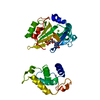
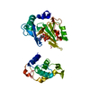
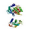
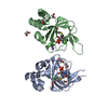
- PDB-6a9w: Structure of the bifunctional DNA primase-polymerase from phage NrS-1 -
+ Open data

- Basic information
| Entry | Database: PDB / ID: 6a9w | ||||||
|---|---|---|---|---|---|---|---|
| Title | Structure of the bifunctional DNA primase-polymerase from phage NrS-1 | ||||||
 Components Components | Primase | ||||||
 Keywords Keywords | REPLICATION / prim-pol | ||||||
| Function / homology |  Function and homology information Function and homology informationviral DNA genome replication / helicase activity / Transferases; Transferring phosphorus-containing groups; Nucleotidyltransferases / transferase activity / DNA helicase / DNA replication / DNA-directed DNA polymerase / hydrolase activity / ATP bindingSimilarity search - Function | ||||||
| Biological species |  Nitratiruptor phage NrS-1 (virus) Nitratiruptor phage NrS-1 (virus) | ||||||
| Method | X-RAY DIFFRACTION / SYNCHROTRON / MOLECULAR REPLACEMENT / molecular replacement / Resolution: 1.8 Å | ||||||
 Authors Authors | Guo, H.J. / Li, M.J. / Wang, T.L. / Wu, H. / Zhou, H. / Xu, C.Y. / Liu, X.P. / Yu, F. / He, J.H. | ||||||
| Funding support |  China, 1items China, 1items
| ||||||
 Citation Citation | Journal: Biochem. Biophys. Res. Commun. / Year: 2019 Title: Crystal structure and biochemical studies of the bifunctional DNA primase-polymerase from phage NrS-1. Authors: Guo, H.J. / Li, M.J. / Wang, T.L. / Wu, H. / Zhou, H. / Xu, C.Y. / Liu, X.P. / Yu, F. / He, J.H. | ||||||
| History |
|
- Structure visualization
| Structure viewer | Molecule: MolmilJmol/JSmol |
|---|
- Downloads & links
-Download
| PDBx/mmCIF format | 6a9w.cif.gz | 131.3 KB | Display | PDBx/mmCIF format |
|---|---|---|---|---|
| PDB format | pdb6a9w.ent.gz | 101.2 KB | Display | PDB format |
| PDBx/mmJSON format | 6a9w.json.gz | Tree view | PDBx/mmJSON format | |
| Others |  Other downloads Other downloads |
-Validation report
| Arichive directory | https://data.pdbj.org/pub/pdb/validation_reports/a9/6a9wftp://data.pdbj.org/pub/pdb/validation_reports/a9/6a9w | HTTPS FTP |
|---|
-Related structure data
| Similar structure data |
|---|


-Links
 PDBj
PDBj- Assembly
| Deposited unit | 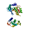
| ||||||||
|---|---|---|---|---|---|---|---|---|---|
| 1 |
| ||||||||
| Unit cell |
|
-Components
| #1: Protein | Mass: 36314.852 Da / Num. of mol.: 1 Source method: isolated from a genetically manipulated source Source: (gene. exp.) Nitratiruptor phage NrS-1 (virus) / Production host:  Escherichia coli (E. coli) / References: UniProt: M5AAG8 Escherichia coli (E. coli) / References: UniProt: M5AAG8 |
|---|---|
| #2: Water | ChemComp-HOH / Water Mass: 18.015 Da / Num. of mol.: 158 / Source method: isolated from a natural source / Formula: H2O Mass: 18.015 Da / Num. of mol.: 158 / Source method: isolated from a natural source / Formula: H2O |
-Experimental details
-Experiment
| Experiment | Method: X-RAY DIFFRACTION / Number of used crystals: 1 |
|---|
- Sample preparation
| Crystal | Density Matthews: 2.26 Å3/Da / Density % sol: 45.51 % |
|---|---|
| Crystal grow | Temperature: 291 K / Method: vapor diffusion, hanging drop / pH: 6.5 / Details: 100mM Bis-Tris, pH 6.5, 0.2M Li2SO4, 25% PEG 3350 |
-Data collection
| Diffraction | Mean temperature: 100 K | ||||||||||||||||||||||||
|---|---|---|---|---|---|---|---|---|---|---|---|---|---|---|---|---|---|---|---|---|---|---|---|---|---|
| Diffraction source | Source: SYNCHROTRON / Site: SSRF / Beamline: BL17U1 / Wavelength: 0.97915 Å | ||||||||||||||||||||||||
| Detector | Type: ADSC QUANTUM 315r / Detector: CCD / Date: Jun 26, 2017 | ||||||||||||||||||||||||
| Radiation | Protocol: SINGLE WAVELENGTH / Monochromatic (M) / Laue (L): M / Scattering type: x-ray | ||||||||||||||||||||||||
| Radiation wavelength | Wavelength: 0.97915 Å / Relative weight: 1 | ||||||||||||||||||||||||
| Reflection | Resolution: 1.8→38.54 Å / Num. obs: 30021 / % possible obs: 99.7 % / Redundancy: 36.8 % / Biso Wilson estimate: 26.13 Å2 / CC1/2: 1 / Rmerge(I) obs: 0.077 / Rpim(I) all: 0.013 / Rrim(I) all: 0.078 / Net I/σ(I): 32.8 | ||||||||||||||||||||||||
| Reflection shell | Diffraction-ID: 1
|
-Phasing
| Phasing | Method: molecular replacement |
|---|
- Processing
| Software |
| ||||||||||||||||||||||||||||||||||||||||||||||||||||||||||||||||||||||||||||||||||||||||||||||||||||
|---|---|---|---|---|---|---|---|---|---|---|---|---|---|---|---|---|---|---|---|---|---|---|---|---|---|---|---|---|---|---|---|---|---|---|---|---|---|---|---|---|---|---|---|---|---|---|---|---|---|---|---|---|---|---|---|---|---|---|---|---|---|---|---|---|---|---|---|---|---|---|---|---|---|---|---|---|---|---|---|---|---|---|---|---|---|---|---|---|---|---|---|---|---|---|---|---|---|---|---|---|---|
| Refinement | Method to determine structure: MOLECULAR REPLACEMENT / Resolution: 1.8→38.54 Å / SU ML: 0.17 / Cross valid method: THROUGHOUT / σ(F): 1.33 / Phase error: 24.37
| ||||||||||||||||||||||||||||||||||||||||||||||||||||||||||||||||||||||||||||||||||||||||||||||||||||
| Solvent computation | Shrinkage radii: 0.9 Å / VDW probe radii: 1.11 Å | ||||||||||||||||||||||||||||||||||||||||||||||||||||||||||||||||||||||||||||||||||||||||||||||||||||
| Displacement parameters | Biso max: 96.33 Å2 / Biso mean: 38.0954 Å2 / Biso min: 16.75 Å2 | ||||||||||||||||||||||||||||||||||||||||||||||||||||||||||||||||||||||||||||||||||||||||||||||||||||
| Refinement step | Cycle: final / Resolution: 1.8→38.54 Å
| ||||||||||||||||||||||||||||||||||||||||||||||||||||||||||||||||||||||||||||||||||||||||||||||||||||
| LS refinement shell | Refine-ID: X-RAY DIFFRACTION / Rfactor Rfree error: 0 / Total num. of bins used: 11
| ||||||||||||||||||||||||||||||||||||||||||||||||||||||||||||||||||||||||||||||||||||||||||||||||||||
| Refinement TLS params. | Method: refined / Refine-ID: X-RAY DIFFRACTION
| ||||||||||||||||||||||||||||||||||||||||||||||||||||||||||||||||||||||||||||||||||||||||||||||||||||
| Refinement TLS group |
|
