 Movie
Movie Controller

[English] 日本語
 Yorodumi
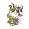
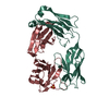
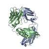
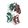
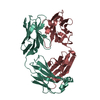
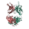
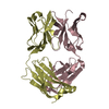
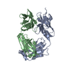
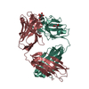
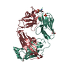
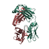
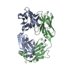
- PDB-5ado: Crystal structure of the paraoxon-modified A.17 antibody FAB frag... -
+ Open data

- Basic information
| Entry | Database: PDB / ID: 5ado | ||||||
|---|---|---|---|---|---|---|---|
| Title | Crystal structure of the paraoxon-modified A.17 antibody FAB fragment - Light chain S35R mutant | ||||||
 Components Components | (FAB A.17) x 2 | ||||||
 Keywords Keywords |  IMMUNE SYSTEM / ENZYME REPROGRAMMING / ANTIBODIES / IG SUPERFAMILY / IN SILICO ANTIBODY MATURATION IMMUNE SYSTEM / ENZYME REPROGRAMMING / ANTIBODIES / IG SUPERFAMILY / IN SILICO ANTIBODY MATURATION | ||||||
| Function / homology | Immunoglobulins / Immunoglobulin-like / Sandwich / Mainly Beta / DIETHYL PHOSPHONATE Function and homology information Function and homology information | ||||||
| Biological species |  Homo sapiens (human) Homo sapiens (human) | ||||||
| Method | X-RAY DIFFRACTION / SYNCHROTRON / MOLECULAR REPLACEMENT / Resolution: 1.55 Å | ||||||
 Authors Authors | Chatziefthimiou, S.D. / Smirnov, I.V. / Golovin, A.V. / Stepanova, A.V. / Peng, Y. / Zolotareva, O.I. / Belogurov, A.A. / Ponomarenko, N.A. / Blackburn, G.M. / Gabibov, A.A. ...Chatziefthimiou, S.D. / Smirnov, I.V. / Golovin, A.V. / Stepanova, A.V. / Peng, Y. / Zolotareva, O.I. / Belogurov, A.A. / Ponomarenko, N.A. / Blackburn, G.M. / Gabibov, A.A. / Lerner, R. / Wilmanns, M. | ||||||
 Citation Citation | Journal: Sci.Adv. / Year: 2016 Title: Robotic Qm/Mm-Driven Maturation of Antibody Combining Sites. Authors: Smirnov, I.V. / Golovin, A.V. / Chatziefthimiou, S.D. / Stepanova, A.V. / Peng, Y. / Zolotareva, O.I. / Belogurov, A.A. / Kurkova, I.N. / Ponomarenko, N.A. / Wilmanns, M. / Blackburn, G.M. / ...Authors: Smirnov, I.V. / Golovin, A.V. / Chatziefthimiou, S.D. / Stepanova, A.V. / Peng, Y. / Zolotareva, O.I. / Belogurov, A.A. / Kurkova, I.N. / Ponomarenko, N.A. / Wilmanns, M. / Blackburn, G.M. / Gabibov, A.G. / Lerner, R.A. | ||||||
| History |
|
- Structure visualization
| Structure viewer | Molecule: MolmilJmol/JSmol |
|---|
- Downloads & links
-Download
| PDBx/mmCIF format | 5ado.cif.gz | 188 KB | Display | PDBx/mmCIF format |
|---|---|---|---|---|
| PDB format | pdb5ado.ent.gz | 148.9 KB | Display | PDB format |
| PDBx/mmJSON format | 5ado.json.gz | Tree view | PDBx/mmJSON format | |
| Others |  Other downloads Other downloads |
-Validation report
| Arichive directory | https://data.pdbj.org/pub/pdb/validation_reports/ad/5adoftp://data.pdbj.org/pub/pdb/validation_reports/ad/5ado | HTTPS FTP |
|---|
-Related structure data
| Related structure data |  5adpC 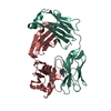 2xzaS C: citing same article ( S: Starting model for refinement |
|---|---|
| Similar structure data |


-Links
 PDBj
PDBj

- Assembly
| Deposited unit | 
| ||||||||
|---|---|---|---|---|---|---|---|---|---|
| 1 |
| ||||||||
| Unit cell |
|
-Components
| #1: Antibody | Mass: 27417.242 Da / Num. of mol.: 1 / Fragment: HEAVY CHAIN Source method: isolated from a genetically manipulated source Source: (gene. exp.) Homo sapiens (human) / Plasmid: PPICZALPHA JK1 / Production host:  KOMAGATAELLA PASTORIS (fungus) / Strain (production host): GS115 KOMAGATAELLA PASTORIS (fungus) / Strain (production host): GS115 | ||
|---|---|---|---|
| #2: Antibody | Mass: 26660.377 Da / Num. of mol.: 1 / Fragment: LIGHT CHAIN Source method: isolated from a genetically manipulated source Source: (gene. exp.) Homo sapiens (human) / Plasmid: PPICZALPHA JK1 / Production host: KOMAGATAELLA PASTORIS (fungus) / Strain (production host): GS115 | ||
| #3: Chemical | ChemComp-DEP /   Mass: 138.102 Da / Num. of mol.: 1 / Source method: obtained synthetically / Formula: C4H11O3P Mass: 138.102 Da / Num. of mol.: 1 / Source method: obtained synthetically / Formula: C4H11O3P | ||
| #4: Water | ChemComp-HOH / Water Mass: 18.015 Da / Num. of mol.: 494 / Source method: isolated from a natural source / Formula: H2O Mass: 18.015 Da / Num. of mol.: 494 / Source method: isolated from a natural source / Formula: H2O | ||
| Nonpolymer details | DIETHYL PHOSPHONAT| Sequence details | CHAIN L HAS MUTATION S35R | |
-Experimental details
-Experiment
| Experiment | Method: X-RAY DIFFRACTION / Number of used crystals: 1 |
|---|
- Sample preparation
| Crystal | Density Matthews: 2.2 Å3/Da / Density % sol: 45 % / Description: NONE |
|---|---|
| Crystal grow | pH: 6.5 Details: 0.18 M MGCL2 16% (W/V) POLYETHYLENE GLYCOL 6000 0.1 M N-(2-ACETAMIDO) IMINODIACETIC ACID (ADA) PH 7.5 |
-Data collection
| Diffraction | Mean temperature: 100 K |
|---|---|
| Diffraction source | Source: SYNCHROTRON / Site: PETRA III, EMBL c/o DESY  / Beamline: P14 (MX2) / Wavelength: 0.8266 / Beamline: P14 (MX2) / Wavelength: 0.8266 |
| Detector | Type: DECTRIS PILATUS 6M / Detector: PIXEL / Date: Dec 6, 2012 / Details: MIRRORS |
| Radiation | Monochromator: SI(III) CRYSTAL MONOCHROMATOR / Protocol: SINGLE WAVELENGTH / Monochromatic (M) / Laue (L): M / Scattering type: x-ray |
| Radiation wavelength | Wavelength: 0.8266 Å / Relative weight: 1 |
| Reflection | Resolution: 1.55→40 Å / Num. obs: 64020 / % possible obs: 99.7 % / Observed criterion σ(I): 2 / Redundancy: 6.7 % / Biso Wilson estimate: 23.5 Å2 / Rmerge(I) obs: 0.05 / Net I/σ(I): 18.5 |
| Reflection shell | Resolution: 1.55→1.65 Å / Redundancy: 6.9 % / Rmerge(I) obs: 0.93 / Mean I/σ(I) obs: 2 / % possible all: 99.8 |
- Processing
| Software |
| ||||||||||||||||||||||||||||||||||||||||||||||||||||||||||||||||||||||||||||||||||||||||||||||||||||||||||||||||||||||||||||||||||||||||||||||||||||||||||||||||||||||||
|---|---|---|---|---|---|---|---|---|---|---|---|---|---|---|---|---|---|---|---|---|---|---|---|---|---|---|---|---|---|---|---|---|---|---|---|---|---|---|---|---|---|---|---|---|---|---|---|---|---|---|---|---|---|---|---|---|---|---|---|---|---|---|---|---|---|---|---|---|---|---|---|---|---|---|---|---|---|---|---|---|---|---|---|---|---|---|---|---|---|---|---|---|---|---|---|---|---|---|---|---|---|---|---|---|---|---|---|---|---|---|---|---|---|---|---|---|---|---|---|---|---|---|---|---|---|---|---|---|---|---|---|---|---|---|---|---|---|---|---|---|---|---|---|---|---|---|---|---|---|---|---|---|---|---|---|---|---|---|---|---|---|---|---|---|---|---|---|---|---|
| Refinement | Method to determine structure: MOLECULAR REPLACEMENT Starting model: PDB ENTRY 2XZA Resolution: 1.55→40 Å / SU ML: 0.19 / σ(F): 2 / Phase error: 21.38 / Stereochemistry target values: ML
| ||||||||||||||||||||||||||||||||||||||||||||||||||||||||||||||||||||||||||||||||||||||||||||||||||||||||||||||||||||||||||||||||||||||||||||||||||||||||||||||||||||||||
| Solvent computation | Shrinkage radii: 0.9 Å / VDW probe radii: 1.11 Å / Solvent model: FLAT BULK SOLVENT MODEL | ||||||||||||||||||||||||||||||||||||||||||||||||||||||||||||||||||||||||||||||||||||||||||||||||||||||||||||||||||||||||||||||||||||||||||||||||||||||||||||||||||||||||
| Displacement parameters | Biso mean: 32 Å2 | ||||||||||||||||||||||||||||||||||||||||||||||||||||||||||||||||||||||||||||||||||||||||||||||||||||||||||||||||||||||||||||||||||||||||||||||||||||||||||||||||||||||||
| Refinement step | Cycle: LAST / Resolution: 1.55→40 Å
| ||||||||||||||||||||||||||||||||||||||||||||||||||||||||||||||||||||||||||||||||||||||||||||||||||||||||||||||||||||||||||||||||||||||||||||||||||||||||||||||||||||||||
| Refine LS restraints |
| ||||||||||||||||||||||||||||||||||||||||||||||||||||||||||||||||||||||||||||||||||||||||||||||||||||||||||||||||||||||||||||||||||||||||||||||||||||||||||||||||||||||||
| LS refinement shell |
| ||||||||||||||||||||||||||||||||||||||||||||||||||||||||||||||||||||||||||||||||||||||||||||||||||||||||||||||||||||||||||||||||||||||||||||||||||||||||||||||||||||||||
| Refinement TLS params. | Method: refined / Refine-ID: X-RAY DIFFRACTION
| ||||||||||||||||||||||||||||||||||||||||||||||||||||||||||||||||||||||||||||||||||||||||||||||||||||||||||||||||||||||||||||||||||||||||||||||||||||||||||||||||||||||||
| Refinement TLS group |
|
