 Movie
Movie Controller

[English] 日本語
 Yorodumi
- PDB-4trp: Crystal structure of monoclonal antibody against neuroblastoma as... -
+ Open data

- Basic information
| Entry | Database: PDB / ID: 4trp | ||||||
|---|---|---|---|---|---|---|---|
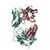
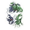
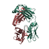
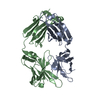
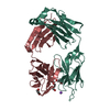
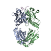
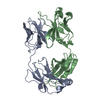
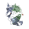
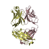
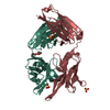
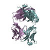
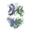
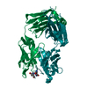
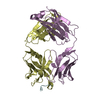
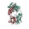
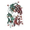
| Title | Crystal structure of monoclonal antibody against neuroblastoma associated antigen. | ||||||
 Components Components |
| ||||||
 Keywords Keywords |  IMMUNE SYSTEM / antibody IMMUNE SYSTEM / antibody | ||||||
| Function / homology | Immunoglobulins / Immunoglobulin-like / Sandwich / Mainly Beta Function and homology information Function and homology information | ||||||
| Biological species |  Mus musculus (house mouse) Mus musculus (house mouse) | ||||||
| Method | X-RAY DIFFRACTION / SYNCHROTRON / Resolution: 1.25 Å | ||||||
 Authors Authors | Horwacik, I. / Golik, P. / Grudnik, P. / Zdzalik, M. / Rokita, H. / Dubin, G. | ||||||
| Funding support |  Poland, 1items Poland, 1items
| ||||||
 Citation Citation | Journal: Mol.Cell Proteomics / Year: 2015 Title: Structural Basis of GD2 Ganglioside and Mimetic Peptide Recognition by 14G2a Antibody. Authors: Horwacik, I. / Golik, P. / Grudnik, P. / Kolinski, M. / Zdzalik, M. / Rokita, H. / Dubin, G. | ||||||
| History |
|
- Structure visualization
| Structure viewer | Molecule: MolmilJmol/JSmol |
|---|
- Downloads & links
-Download
| PDBx/mmCIF format | 4trp.cif.gz | 189.4 KB | Display | PDBx/mmCIF format |
|---|---|---|---|---|
| PDB format | pdb4trp.ent.gz | 148.5 KB | Display | PDB format |
| PDBx/mmJSON format | 4trp.json.gz | Tree view | PDBx/mmJSON format | |
| Others |  Other downloads Other downloads |
-Validation report
| Arichive directory | https://data.pdbj.org/pub/pdb/validation_reports/tr/4trpftp://data.pdbj.org/pub/pdb/validation_reports/tr/4trp | HTTPS FTP |
|---|
-Related structure data
| Related structure data |  4tujC  4tukC  4tulC  4tuoC 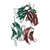 1f8tS S: Starting model for refinement C: citing same article ( |
|---|---|
| Similar structure data |


-Links
 PDBj
PDBj

- Assembly
| Deposited unit | 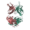
| ||||||||
|---|---|---|---|---|---|---|---|---|---|
| 1 |
| ||||||||
| Unit cell |
|
-Components
| #1: Antibody | Mass: 22748.438 Da / Num. of mol.: 1 Source method: isolated from a genetically manipulated source Source: (gene. exp.) Mus musculus (house mouse) / Production host: Mus musculus (house mouse) |
|---|---|
| #2: Antibody | Mass: 24251.980 Da / Num. of mol.: 1 Source method: isolated from a genetically manipulated source Source: (gene. exp.) Mus musculus (house mouse) / Production host: Mus musculus (house mouse) |
| #3: Water | ChemComp-HOH / Water Mass: 18.015 Da / Num. of mol.: 525 / Source method: isolated from a natural source / Formula: H2O Mass: 18.015 Da / Num. of mol.: 525 / Source method: isolated from a natural source / Formula: H2O |
-Experimental details
-Experiment
| Experiment | Method: X-RAY DIFFRACTION |
|---|
- Sample preparation
| Crystal | Density Matthews: 2.44 Å3/Da / Density % sol: 49.61 % |
|---|---|
| Crystal grow | Temperature: 293 K / Method: vapor diffusion, sitting drop / Details: Hepes, PEG 4000, isopropanol |
-Data collection
| Diffraction | Mean temperature: 80 K |
|---|---|
| Diffraction source | Source: SYNCHROTRON / Site: PETRA III, EMBL c/o DESY  / Beamline: P13 (MX1) / Wavelength: 0.88563 Å / Beamline: P13 (MX1) / Wavelength: 0.88563 Å |
| Detector | Type: DECTRIS PILATUS 6M / Detector: PIXEL / Date: Dec 10, 2013 |
| Radiation | Protocol: SINGLE WAVELENGTH / Monochromatic (M) / Laue (L): M / Scattering type: x-ray |
| Radiation wavelength | Wavelength: 0.88563 Å / Relative weight: 1 |
| Reflection | Resolution: 1.25→57.86 Å / Num. obs: 109662 / % possible obs: 89.14 % / Redundancy: 3.8 % / Net I/σ(I): 10.99 |
| Reflection shell | Resolution: 1.25→1.32 Å / Redundancy: 3.8 % / Rmerge(I) obs: 0.457 / Mean I/σ(I) obs: 2.71 / % possible all: 86.7 |
- Processing
| Software |
| |||||||||||||||||||||||||||||||||||||||||||||||||||||||||||||||||||||||||||||||||||||||||||||||||||||||||||||||||||||||||||||||||||||||||||||||||||||||||||||||||||||||||||||||||||||||||||||||||||||||||||||||||||||||||
|---|---|---|---|---|---|---|---|---|---|---|---|---|---|---|---|---|---|---|---|---|---|---|---|---|---|---|---|---|---|---|---|---|---|---|---|---|---|---|---|---|---|---|---|---|---|---|---|---|---|---|---|---|---|---|---|---|---|---|---|---|---|---|---|---|---|---|---|---|---|---|---|---|---|---|---|---|---|---|---|---|---|---|---|---|---|---|---|---|---|---|---|---|---|---|---|---|---|---|---|---|---|---|---|---|---|---|---|---|---|---|---|---|---|---|---|---|---|---|---|---|---|---|---|---|---|---|---|---|---|---|---|---|---|---|---|---|---|---|---|---|---|---|---|---|---|---|---|---|---|---|---|---|---|---|---|---|---|---|---|---|---|---|---|---|---|---|---|---|---|---|---|---|---|---|---|---|---|---|---|---|---|---|---|---|---|---|---|---|---|---|---|---|---|---|---|---|---|---|---|---|---|---|---|---|---|---|---|---|---|---|---|---|---|---|---|---|---|---|
| Refinement | Starting model: 1f8t Resolution: 1.25→42.914 Å / SU ML: 0.12 / Cross valid method: FREE R-VALUE / σ(F): 1.96 / Phase error: 17.93 / Stereochemistry target values: ML
| |||||||||||||||||||||||||||||||||||||||||||||||||||||||||||||||||||||||||||||||||||||||||||||||||||||||||||||||||||||||||||||||||||||||||||||||||||||||||||||||||||||||||||||||||||||||||||||||||||||||||||||||||||||||||
| Solvent computation | Shrinkage radii: 0.9 Å / VDW probe radii: 1.11 Å / Solvent model: FLAT BULK SOLVENT MODEL | |||||||||||||||||||||||||||||||||||||||||||||||||||||||||||||||||||||||||||||||||||||||||||||||||||||||||||||||||||||||||||||||||||||||||||||||||||||||||||||||||||||||||||||||||||||||||||||||||||||||||||||||||||||||||
| Refinement step | Cycle: LAST / Resolution: 1.25→42.914 Å
| |||||||||||||||||||||||||||||||||||||||||||||||||||||||||||||||||||||||||||||||||||||||||||||||||||||||||||||||||||||||||||||||||||||||||||||||||||||||||||||||||||||||||||||||||||||||||||||||||||||||||||||||||||||||||
| Refine LS restraints |
| |||||||||||||||||||||||||||||||||||||||||||||||||||||||||||||||||||||||||||||||||||||||||||||||||||||||||||||||||||||||||||||||||||||||||||||||||||||||||||||||||||||||||||||||||||||||||||||||||||||||||||||||||||||||||
| LS refinement shell |
|
