 Movie
Movie Controller


+ Open data

- Basic information
| Entry | Database: PDB / ID: 3l7e | ||||||
|---|---|---|---|---|---|---|---|
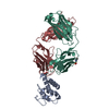
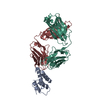
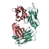
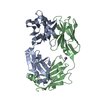
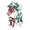
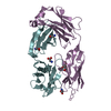
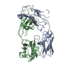
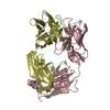
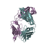
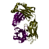
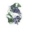
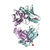
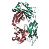
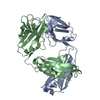
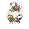
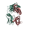
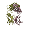
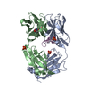
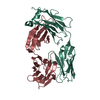
| Title | Crystal structure of ANTI-IL-13 antibody C836 | ||||||
 Components Components |
| ||||||
 Keywords Keywords |  IMMUNE SYSTEM / immunoglobulin fold / MONOCLONAL ANTIBODY IMMUNE SYSTEM / immunoglobulin fold / MONOCLONAL ANTIBODY | ||||||
| Function / homology | Immunoglobulins / Immunoglobulin-like / Sandwich / Mainly Beta / ACETATE ION Function and homology information Function and homology information | ||||||
| Biological species |  Mus musculus (house mouse) Mus musculus (house mouse) Homo sapiens (human) Homo sapiens (human) | ||||||
| Method | X-RAY DIFFRACTION / MOLECULAR REPLACEMENT / Resolution: 2.5 Å | ||||||
 Authors Authors | Teplyakov, A. / Obmolova, G. / Malia, T. / Gilliland, G.L. | ||||||
 Citation Citation | Journal: Acta Crystallogr.,Sect.F / Year: 2011 Title: Antigen recognition by antibody C836 through adjustment of VL/VH packing Authors: Teplyakov, A. / Obmolova, G. / Malia, T.J. / Gilliland, G. | ||||||
| History |
|
- Structure visualization
| Structure viewer | Molecule: MolmilJmol/JSmol |
|---|
- Downloads & links
-Download
| PDBx/mmCIF format | 3l7e.cif.gz | 177.7 KB | Display | PDBx/mmCIF format |
|---|---|---|---|---|
| PDB format | pdb3l7e.ent.gz | 140.2 KB | Display | PDB format |
| PDBx/mmJSON format | 3l7e.json.gz | Tree view | PDBx/mmJSON format | |
| Others |  Other downloads Other downloads |
-Validation report
| Arichive directory | https://data.pdbj.org/pub/pdb/validation_reports/l7/3l7eftp://data.pdbj.org/pub/pdb/validation_reports/l7/3l7e | HTTPS FTP |
|---|
-Related structure data
| Related structure data | 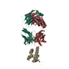 3l5wS S: Starting model for refinement |
|---|---|
| Similar structure data |


-Links
 PDBj
PDBj

- Assembly
| Deposited unit | 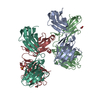
| ||||||||
|---|---|---|---|---|---|---|---|---|---|
| 1 | 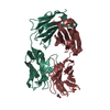
| ||||||||
| 2 | 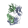
| ||||||||
| Unit cell |
|
-Components
-Antibody , 2 types, 4 molecules LAHB
| #1: Antibody | Mass: 23503.090 Da / Num. of mol.: 2 Fragment: CHIMERIC MOLECULE OF MOUSE VARIABLE DOMAIN AND HUMAN CONSTANT DOMAIN Source method: isolated from a genetically manipulated source Source: (gene. exp.) Mus musculus, Homo sapiens Cell (production host): Human embryonic kidney (HEK) 293 cells Production host: Homo sapiens (human)#2: Antibody | Mass: 24780.824 Da / Num. of mol.: 2 Fragment: FD fragment of the heavy chain, CHIMERIC MOLECULE OF MOUSE VARIABLE DOMAIN AND HUMAN CONSTANT DOMAIN Source method: isolated from a genetically manipulated source Source: (gene. exp.) Mus musculus, Homo sapiens Cell (production host): Human embryonic kidney (HEK) 293 cells Production host: Homo sapiens (human) |
|---|
-Non-polymers , 4 types, 179 molecules 


| #3: Chemical | Sulfate Mass: 96.063 Da / Num. of mol.: 2 / Source method: obtained synthetically / Formula: SO4 Mass: 96.063 Da / Num. of mol.: 2 / Source method: obtained synthetically / Formula: SO4#4: Chemical | ChemComp-GOL / | Glycerol Mass: 92.094 Da / Num. of mol.: 1 / Source method: obtained synthetically / Formula: C3H8O3 Mass: 92.094 Da / Num. of mol.: 1 / Source method: obtained synthetically / Formula: C3H8O3#5: Chemical | Acetate Mass: 59.044 Da / Num. of mol.: 2 / Source method: obtained synthetically / Formula: C2H3O2 Mass: 59.044 Da / Num. of mol.: 2 / Source method: obtained synthetically / Formula: C2H3O2#6: Water | ChemComp-HOH / | WaterMass: 18.015 Da / Num. of mol.: 174 / Source method: isolated from a natural source / Formula: H2O |
|---|
-Experimental details
-Experiment
| Experiment | Method: X-RAY DIFFRACTION / Number of used crystals: 1 |
|---|
- Sample preparation
| Crystal | Density Matthews: 2.44 Å3/Da / Density % sol: 49 % |
|---|---|
| Crystal grow | Temperature: 293 K / Method: vapor diffusion, sitting drop / pH: 4.5 Details: 0.1 M SODIUM ACETATE PH 4.5, 0.2 M AMMONIUM SULFATE, 16% PEG 4K; CRYO CONDITIONS: SODIUM ACETATE PH 4.5, 0.2 M AMMONIUM SULFATE, 20% PEG 4K, 20% GLYCEROL, VAPOR DIFFUSION, SITTING DROP, temperature 293K |
-Data collection
| Diffraction | Mean temperature: 120 K |
|---|---|
| Diffraction source | Source: ROTATING ANODE / Type: RIGAKU MICROMAX-007 HF / Wavelength: 1.5418 / Wavelength: 1.5418 Å |
| Detector | Type: RIGAKU SATURN 944 / Detector: CCD / Date: Jun 11, 2007 / Details: VARIMAX HF |
| Radiation | Protocol: SINGLE WAVELENGTH / Monochromatic (M) / Laue (L): M / Scattering type: x-ray |
| Radiation wavelength | Wavelength: 1.5418 Å / Relative weight: 1 |
| Reflection | Resolution: 2.5→49 Å / Num. all: 30327 / Num. obs: 30327 / % possible obs: 94.8 % / Observed criterion σ(I): -3 / Redundancy: 7.7 % / Biso Wilson estimate: 59.8 Å2 / Rmerge(I) obs: 0.08 / Net I/σ(I): 10.4 |
| Reflection shell | Resolution: 2.5→2.59 Å / Redundancy: 7 % / Rmerge(I) obs: 0.408 / Mean I/σ(I) obs: 3 / % possible all: 75.7 |
- Processing
| Software |
| ||||||||||||||||||||||||||||||||||||||||||||||||||||||||||||||||||||||||||||||||||||||||||
|---|---|---|---|---|---|---|---|---|---|---|---|---|---|---|---|---|---|---|---|---|---|---|---|---|---|---|---|---|---|---|---|---|---|---|---|---|---|---|---|---|---|---|---|---|---|---|---|---|---|---|---|---|---|---|---|---|---|---|---|---|---|---|---|---|---|---|---|---|---|---|---|---|---|---|---|---|---|---|---|---|---|---|---|---|---|---|---|---|---|---|---|
| Refinement | Method to determine structure: MOLECULAR REPLACEMENT Starting model: PDB ENTRY 3L5W Resolution: 2.5→15 Å / Cor.coef. Fo:Fc: 0.947 / Cor.coef. Fo:Fc free: 0.92 / SU B: 9.348 / SU ML: 0.208 / Cross valid method: THROUGHOUT / σ(F): 0 / ESU R: 1.035 / ESU R Free: 0.31 / Stereochemistry target values: Engh & Huber
| ||||||||||||||||||||||||||||||||||||||||||||||||||||||||||||||||||||||||||||||||||||||||||
| Solvent computation | Ion probe radii: 0.8 Å / Shrinkage radii: 0.8 Å / VDW probe radii: 1.2 Å / Solvent model: BABINET MODEL WITH MASK | ||||||||||||||||||||||||||||||||||||||||||||||||||||||||||||||||||||||||||||||||||||||||||
| Displacement parameters | Biso mean: 45.9 Å2
| ||||||||||||||||||||||||||||||||||||||||||||||||||||||||||||||||||||||||||||||||||||||||||
| Refine analyze | Luzzati coordinate error free: 0.31 Å | ||||||||||||||||||||||||||||||||||||||||||||||||||||||||||||||||||||||||||||||||||||||||||
| Refinement step | Cycle: LAST / Resolution: 2.5→15 Å
| ||||||||||||||||||||||||||||||||||||||||||||||||||||||||||||||||||||||||||||||||||||||||||
| Refine LS restraints |
| ||||||||||||||||||||||||||||||||||||||||||||||||||||||||||||||||||||||||||||||||||||||||||
| LS refinement shell | Resolution: 2.5→2.563 Å / Total num. of bins used: 20
|
