 Movie
Movie Controller

[English] 日本語
 Yorodumi
- PDB-4zcs: Crystal structure of the C-terminal catalytic domain of Plasmodiu... -
+ Open data

- Basic information
| Entry | Database: PDB / ID: 4zcs | ||||||
|---|---|---|---|---|---|---|---|
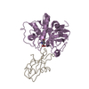
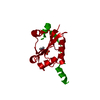
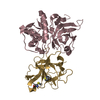
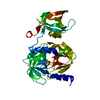
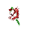
| Title | Crystal structure of the C-terminal catalytic domain of Plasmodium falciparum CTP:phosphocholine cytidylyltransferase in complex with CDP-choline | ||||||
 Components Components | Choline-phosphate cytidylyltransferase | ||||||
 Keywords Keywords | TRANSFERASE / enzyme / malaria / cytidylyltransferase / phosphatidylcholine | ||||||
| Function / homology |  Function and homology information Function and homology informationSynthesis of PC / choline-phosphate cytidylyltransferase / choline-phosphate cytidylyltransferase activity / phosphatidylcholine binding / identical protein bindingSimilarity search - Function | ||||||
| Biological species |  Plasmodium falciparum (malaria parasite P. falciparum) Plasmodium falciparum (malaria parasite P. falciparum) | ||||||
| Method | X-RAY DIFFRACTION / SYNCHROTRON / MOLECULAR REPLACEMENT / Resolution: 2.45 Å | ||||||
 Authors Authors | Guca, E. / Hoh, F. / Guichou, J.-F. / Cerdan, R. | ||||||
| Funding support |  France, 1items France, 1items
| ||||||
 Citation Citation | Journal: Sci Rep / Year: 2018 Title: Structural determinants of the catalytic mechanism of Plasmodium CCT, a key enzyme of malaria lipid biosynthesis. Authors: Guca, E. / Nagy, G.N. / Hajdu, F. / Marton, L. / Izrael, R. / Hoh, F. / Yang, Y. / Vial, H. / Vertessy, B.G. / Guichou, J.F. / Cerdan, R. | ||||||
| History |
|
- Structure visualization
| Structure viewer | Molecule: MolmilJmol/JSmol |
|---|
- Downloads & links
-Download
| PDBx/mmCIF format | 4zcs.cif.gz | 197 KB | Display | PDBx/mmCIF format |
|---|---|---|---|---|
| PDB format | pdb4zcs.ent.gz | 156.6 KB | Display | PDB format |
| PDBx/mmJSON format | 4zcs.json.gz | Tree view | PDBx/mmJSON format | |
| Others |  Other downloads Other downloads |
-Validation report
| Arichive directory | https://data.pdbj.org/pub/pdb/validation_reports/zc/4zcsftp://data.pdbj.org/pub/pdb/validation_reports/zc/4zcs | HTTPS FTP |
|---|
-Related structure data
| Related structure data |  4zcpC  4zcqC  4zcrC  4zctC  3hl4S C: citing same article ( S: Starting model for refinement |
|---|---|
| Similar structure data |


-Links
 PDBj
PDBj- Assembly
| Deposited unit | 
| ||||||||
|---|---|---|---|---|---|---|---|---|---|
| 1 | 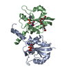
| ||||||||
| 2 | 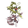
| ||||||||
| 3 | 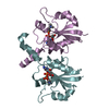
| ||||||||
| Unit cell |
|
-Components
| #1: Protein | Mass: 20810.123 Da / Num. of mol.: 6 / Fragment: UNP residues 581-775 Source method: isolated from a genetically manipulated source Source: (gene. exp.) Plasmodium falciparum (isolate 3D7) (eukaryote)Gene: PF3D7_1316600 / Production host:  Escherichia coli (E. coli) Escherichia coli (E. coli)References: UniProt: Q8IEE9, choline-phosphate cytidylyltransferase#2: Chemical | ChemComp-CDC / [ Citicoline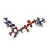  Mass: 488.324 Da / Num. of mol.: 6 / Source method: obtained synthetically / Formula: C14H26N4O11P2 Mass: 488.324 Da / Num. of mol.: 6 / Source method: obtained synthetically / Formula: C14H26N4O11P2#3: Chemical | Polyethylene glycol  Mass: 326.383 Da / Num. of mol.: 2 / Source method: obtained synthetically / Formula: C14H30O8 / Comment: precipitant*YM Mass: 326.383 Da / Num. of mol.: 2 / Source method: obtained synthetically / Formula: C14H30O8 / Comment: precipitant*YM#4: Water | ChemComp-HOH / | Water Mass: 18.015 Da / Num. of mol.: 341 / Source method: isolated from a natural source / Formula: H2O Mass: 18.015 Da / Num. of mol.: 341 / Source method: isolated from a natural source / Formula: H2O |
|---|
-Experimental details
-Experiment
| Experiment | Method: X-RAY DIFFRACTION / Number of used crystals: 1 |
|---|
- Sample preparation
| Crystal | Density Matthews: 3.01 Å3/Da / Density % sol: 59.07 % |
|---|---|
| Crystal grow | Temperature: 293 K / Method: vapor diffusion, sitting drop / pH: 6.9 / Details: 0.2M NaF, 20% PEG 3350 |
-Data collection
| Diffraction | Mean temperature: 100 K |
|---|---|
| Diffraction source | Source: SYNCHROTRON / Site: ESRF / Beamline: ID23-1 / Wavelength: 0.979338 Å |
| Detector | Type: DECTRIS PILATUS 6M-F / Detector: PIXEL / Date: Jan 31, 2014 |
| Radiation | Protocol: SINGLE WAVELENGTH / Monochromatic (M) / Laue (L): M / Scattering type: x-ray |
| Radiation wavelength | Wavelength: 0.979338 Å / Relative weight: 1 |
| Reflection | Resolution: 2.45→113.67 Å / Num. obs: 52521 / % possible obs: 99.63 % / Redundancy: 9.35 % / Rsym value: 0.32 / Net I/σ(I): 0.2 |
- Processing
| Software |
| ||||||||||||||||||||||||||||||||||||||||||||||||||||||||||||||||||||||||||||||||||||||||||||||||||||||||||||||||||||||||||||||||||||||||||||||||||||||||||||||||||||||||||||||||||||||
|---|---|---|---|---|---|---|---|---|---|---|---|---|---|---|---|---|---|---|---|---|---|---|---|---|---|---|---|---|---|---|---|---|---|---|---|---|---|---|---|---|---|---|---|---|---|---|---|---|---|---|---|---|---|---|---|---|---|---|---|---|---|---|---|---|---|---|---|---|---|---|---|---|---|---|---|---|---|---|---|---|---|---|---|---|---|---|---|---|---|---|---|---|---|---|---|---|---|---|---|---|---|---|---|---|---|---|---|---|---|---|---|---|---|---|---|---|---|---|---|---|---|---|---|---|---|---|---|---|---|---|---|---|---|---|---|---|---|---|---|---|---|---|---|---|---|---|---|---|---|---|---|---|---|---|---|---|---|---|---|---|---|---|---|---|---|---|---|---|---|---|---|---|---|---|---|---|---|---|---|---|---|---|---|
| Refinement | Method to determine structure: MOLECULAR REPLACEMENT Starting model: 3HL4 Resolution: 2.45→113.67 Å / Cor.coef. Fo:Fc: 0.956 / Cor.coef. Fo:Fc free: 0.921 / SU B: 7.35 / SU ML: 0.162 / Cross valid method: THROUGHOUT / ESU R: 0.255 / ESU R Free: 0.219 / Stereochemistry target values: MAXIMUM LIKELIHOOD / Details: HYDROGENS HAVE BEEN ADDED IN THE RIDING POSITIONS
| ||||||||||||||||||||||||||||||||||||||||||||||||||||||||||||||||||||||||||||||||||||||||||||||||||||||||||||||||||||||||||||||||||||||||||||||||||||||||||||||||||||||||||||||||||||||
| Solvent computation | Ion probe radii: 0.8 Å / Shrinkage radii: 0.8 Å / VDW probe radii: 1.2 Å / Solvent model: MASK | ||||||||||||||||||||||||||||||||||||||||||||||||||||||||||||||||||||||||||||||||||||||||||||||||||||||||||||||||||||||||||||||||||||||||||||||||||||||||||||||||||||||||||||||||||||||
| Displacement parameters | Biso mean: 44.051 Å2
| ||||||||||||||||||||||||||||||||||||||||||||||||||||||||||||||||||||||||||||||||||||||||||||||||||||||||||||||||||||||||||||||||||||||||||||||||||||||||||||||||||||||||||||||||||||||
| Refinement step | Cycle: LAST / Resolution: 2.45→113.67 Å
| ||||||||||||||||||||||||||||||||||||||||||||||||||||||||||||||||||||||||||||||||||||||||||||||||||||||||||||||||||||||||||||||||||||||||||||||||||||||||||||||||||||||||||||||||||||||
| Refine LS restraints |
|