 Movie
Movie Controller

[English] 日本語
 Yorodumi
- PDB-3hl4: Crystal structure of a mammalian CTP:phosphocholine cytidylyltran... -
+ Open data

- Basic information
| Entry | Database: PDB / ID: 3hl4 | ||||||
|---|---|---|---|---|---|---|---|
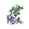
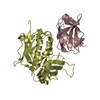
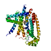
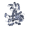
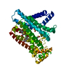
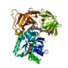
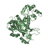
| Title | Crystal structure of a mammalian CTP:phosphocholine cytidylyltransferase with CDP-choline | ||||||
 Components Components | Choline-phosphate cytidylyltransferase A | ||||||
 Keywords Keywords |  TRANSFERASE / Cytidylyltransferase / Rossmann Fold / Phospholipid synthesis / Phosphatidylcholine / Phosphocholine / CTP / CDP-choline / Amphitropic protein / Lipid metabolism / Nucleotidyltransferase / Phosphoprotein TRANSFERASE / Cytidylyltransferase / Rossmann Fold / Phospholipid synthesis / Phosphatidylcholine / Phosphocholine / CTP / CDP-choline / Amphitropic protein / Lipid metabolism / Nucleotidyltransferase / Phosphoprotein | ||||||
| Function / homology |  Function and homology information Function and homology informationSynthesis of PC / choline-phosphate cytidylyltransferase / choline-phosphate cytidylyltransferase activity / CDP-choline pathway / glycogen granule / phosphatidylcholine biosynthetic process / phosphatidylcholine binding / extrinsic component of membrane / molecular function inhibitor activity / nuclear envelope ...Synthesis of PC / choline-phosphate cytidylyltransferase / choline-phosphate cytidylyltransferase activity / CDP-choline pathway / glycogen granule / phosphatidylcholine biosynthetic process / phosphatidylcholine binding / extrinsic component of membrane / molecular function inhibitor activity / nuclear envelope / calmodulin binding / lipid binding / endoplasmic reticulum membrane / endoplasmic reticulum / protein homodimerization activity / identical protein binding / nucleus / cytosolSimilarity search - Function | ||||||
| Biological species |  Rattus norvegicus (Norway rat) Rattus norvegicus (Norway rat) | ||||||
| Method | X-RAY DIFFRACTION / SYNCHROTRON / SAD / Resolution: 2.2 Å | ||||||
 Authors Authors | Lee, J. / Paetzel, M. / Cornell, R.B. | ||||||
 Citation Citation | Journal: J.Biol.Chem. / Year: 2009 Title: Crystal Structure of a mammalian CTP: Phosphocholine cytidylyltransferase catalytic domain reveals novel active site residues within a highly conserved nucleotidyl-transferase fold Authors: Lee, J. / Johnson, J.E. / Ding, Z. / Paetzel, M. / Cornell, R.B. | ||||||
| History |
|
- Structure visualization
| Structure viewer | Molecule: MolmilJmol/JSmol |
|---|
- Downloads & links
-Download
| PDBx/mmCIF format | 3hl4.cif.gz | 92.5 KB | Display | PDBx/mmCIF format |
|---|---|---|---|---|
| PDB format | pdb3hl4.ent.gz | 70.2 KB | Display | PDB format |
| PDBx/mmJSON format | 3hl4.json.gz | Tree view | PDBx/mmJSON format | |
| Others |  Other downloads Other downloads |
-Validation report
| Arichive directory | https://data.pdbj.org/pub/pdb/validation_reports/hl/3hl4ftp://data.pdbj.org/pub/pdb/validation_reports/hl/3hl4 | HTTPS FTP |
|---|
-Related structure data
| Similar structure data |
|---|


-Links
 PDBj
PDBj- Assembly
| Deposited unit | 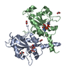
| ||||||||
|---|---|---|---|---|---|---|---|---|---|
| 1 |
| ||||||||
| Unit cell |
| ||||||||
| Details | Biological unit is a dimer in ASU |
-Components
| #1: Protein | Mass: 26792.066 Da / Num. of mol.: 2 / Fragment: Domain N and C / Mutation: None Source method: isolated from a genetically manipulated source Source: (gene. exp.) Rattus norvegicus (Norway rat) / Gene: Ctpct, Pcyt1, Pcyt1a / Plasmid: PVL1393 / Production host:  TRICHOPLUSIA NI (cabbage looper) TRICHOPLUSIA NI (cabbage looper)References: UniProt: P19836, choline-phosphate cytidylyltransferase#2: Chemical | Citicoline  Mass: 488.324 Da / Num. of mol.: 2 / Source method: obtained synthetically / Formula: C14H26N4O11P2 Mass: 488.324 Da / Num. of mol.: 2 / Source method: obtained synthetically / Formula: C14H26N4O11P2#3: Chemical | ChemComp-GOL / | Glycerol  Mass: 92.094 Da / Num. of mol.: 1 / Source method: obtained synthetically / Formula: C3H8O3 Mass: 92.094 Da / Num. of mol.: 1 / Source method: obtained synthetically / Formula: C3H8O3#4: Chemical | ChemComp-FMT / Formic acid  Mass: 46.025 Da / Num. of mol.: 7 / Source method: obtained synthetically / Formula: CH2O2 Mass: 46.025 Da / Num. of mol.: 7 / Source method: obtained synthetically / Formula: CH2O2#5: Water | ChemComp-HOH / | Water Mass: 18.015 Da / Num. of mol.: 249 / Source method: isolated from a natural source / Formula: H2O Mass: 18.015 Da / Num. of mol.: 249 / Source method: isolated from a natural source / Formula: H2O |
|---|
-Experimental details
-Experiment
| Experiment | Method: X-RAY DIFFRACTION / Number of used crystals: 1 |
|---|
- Sample preparation
| Crystal | Density Matthews: 2.34 Å3/Da / Density % sol: 47.42 % |
|---|---|
| Crystal grow | Temperature: 291 K / Method: vapor diffusion, sitting drop / pH: 4.6 Details: 0.1M Na Acetate 1.4M Na Formate, pH 4.6, VAPOR DIFFUSION, SITTING DROP, temperature 291K |
-Data collection
| Diffraction | Mean temperature: 100 K |
|---|---|
| Diffraction source | Source: SYNCHROTRON / Site: ALS  / Beamline: 8.2.2 / Wavelength: 1 Å / Beamline: 8.2.2 / Wavelength: 1 Å |
| Detector | Type: ADSC QUANTUM 315 / Detector: CCD / Date: Jul 7, 2005 / Details: KOHZU: Double Crystal Si(111) |
| Radiation | Monochromator: KOHZU / Protocol: SINGLE WAVELENGTH / Monochromatic (M) / Laue (L): M / Scattering type: x-ray |
| Radiation wavelength | Wavelength: 1 Å / Relative weight: 1 |
| Reflection | Resolution: 2.2→50 Å / Num. all: 26316 / Num. obs: 26316 / % possible obs: 100 % / Redundancy: 11.2 % / Rmerge(I) obs: 0.11 / Net I/σ(I): 32.7 |
| Reflection shell | Resolution: 2.2→2.28 Å / Redundancy: 10.6 % / Rmerge(I) obs: 0.33 / Mean I/σ(I) obs: 6.6 / Num. unique all: 2587 / % possible all: 100 |
- Processing
| Software |
| |||||||||||||||||||||||||||||||||||||||||||||||||||||||||||||||||
|---|---|---|---|---|---|---|---|---|---|---|---|---|---|---|---|---|---|---|---|---|---|---|---|---|---|---|---|---|---|---|---|---|---|---|---|---|---|---|---|---|---|---|---|---|---|---|---|---|---|---|---|---|---|---|---|---|---|---|---|---|---|---|---|---|---|---|
| Refinement | Method to determine structure: SAD / Resolution: 2.2→50 Å / Cor.coef. Fo:Fc: 0.949 / Cor.coef. Fo:Fc free: 0.937 / SU B: 4.862 / SU ML: 0.129 / Cross valid method: THROUGHOUT / ESU R: 0.252 / ESU R Free: 0.213 / Stereochemistry target values: MAXIMUM LIKELIHOOD / Details: HYDROGENS HAVE BEEN ADDED IN THE RIDING POSITIONS
| |||||||||||||||||||||||||||||||||||||||||||||||||||||||||||||||||
| Solvent computation | Ion probe radii: 0.8 Å / Shrinkage radii: 0.8 Å / VDW probe radii: 1.4 Å / Solvent model: MASK | |||||||||||||||||||||||||||||||||||||||||||||||||||||||||||||||||
| Displacement parameters | Biso mean: 27.857 Å2
| |||||||||||||||||||||||||||||||||||||||||||||||||||||||||||||||||
| Refinement step | Cycle: LAST / Resolution: 2.2→50 Å
| |||||||||||||||||||||||||||||||||||||||||||||||||||||||||||||||||
| Refine LS restraints |
| |||||||||||||||||||||||||||||||||||||||||||||||||||||||||||||||||
| LS refinement shell | Resolution: 2.201→2.258 Å / Total num. of bins used: 20
|
