 Movie
Movie Controller

[English] 日本語
 Yorodumi
- PDB-4z2q: The crystal structure of Sclerotium Rolfsii lectin variant 1 (SSR... -
+ Open data

- Basic information
| Entry | Database: PDB / ID: 4z2q | |||||||||
|---|---|---|---|---|---|---|---|---|---|---|
| Title | The crystal structure of Sclerotium Rolfsii lectin variant 1 (SSR1) in complex with N-acetyl-glucosamine | |||||||||
 Components Components | Lectin | |||||||||
 Keywords Keywords | SUGAR BINDING PROTEIN / Lectin / carbohydrate- binding specificity | |||||||||
| Function / homology | Fungal fruit body lectin / Fungal fruit body lectin / Cytolysin/lectin / Cytolysin/lectin / Mutm (Fpg) Protein; Chain: A, domain 2 / Sandwich / Mainly Beta / 2-acetamido-2-deoxy-alpha-D-glucopyranose / Lectin Function and homology information Function and homology information | |||||||||
| Biological species |  Athelia rolfsii (fungus) Athelia rolfsii (fungus) | |||||||||
| Method | X-RAY DIFFRACTION / MOLECULAR REPLACEMENT / Resolution: 1.9 Å | |||||||||
 Authors Authors | Kantsadi, A.L. / Peppa, V.I. / Leonidas, D.D. | |||||||||
 Citation Citation | Journal: Molecules / Year: 2015 Title: Molecular Cloning, Carbohydrate Specificity and the Crystal Structure of Two Sclerotium rolfsii Lectin Variants. Authors: Peppa, V.I. / Venkat, H. / Kantsadi, A.L. / Inamdar, S.R. / Bhat, G.G. / Eligar, S. / Shivanand, A. / Chachadi, V.B. / Satisha, G.J. / Swamy, B.M. / Skamnaki, V.T. / Zographos, S.E. / Leonidas, D.D. | |||||||||
| History |
|
- Structure visualization
| Structure viewer | Molecule: MolmilJmol/JSmol |
|---|
- Downloads & links
-Download
| PDBx/mmCIF format | 4z2q.cif.gz | 80.3 KB | Display | PDBx/mmCIF format |
|---|---|---|---|---|
| PDB format | pdb4z2q.ent.gz | 59.2 KB | Display | PDB format |
| PDBx/mmJSON format | 4z2q.json.gz | Tree view | PDBx/mmJSON format | |
| Others |  Other downloads Other downloads |
-Validation report
| Arichive directory | https://data.pdbj.org/pub/pdb/validation_reports/z2/4z2qftp://data.pdbj.org/pub/pdb/validation_reports/z2/4z2q | HTTPS FTP |
|---|
-Related structure data
| Related structure data | 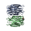 4yldC 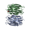 4z2fC 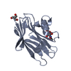 4z2sC 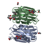 2ofeS S: Starting model for refinement C: citing same article ( |
|---|---|
| Similar structure data |


-Links
 PDBj
PDBj


- Assembly
| Deposited unit | 
| |||||||||
|---|---|---|---|---|---|---|---|---|---|---|
| 1 |
| |||||||||
| Unit cell |
| |||||||||
| Components on special symmetry positions |
|
-Components
-Protein , 1 types, 2 molecules AB
| #1: Protein | Mass: 16062.690 Da / Num. of mol.: 2 Source method: isolated from a genetically manipulated source Source: (gene. exp.) Athelia rolfsii (fungus) / Gene: l1 / Production host:  Escherichia coli (E. coli) / References: UniProt: A0A0M3KL30*PLUS Escherichia coli (E. coli) / References: UniProt: A0A0M3KL30*PLUS |
|---|
-Sugars , 2 types, 4 molecules 

| #3: Sugar | N-Acetylglucosamine Type: D-saccharide, alpha linking / Mass: 221.208 Da / Num. of mol.: 2 Type: D-saccharide, alpha linking / Mass: 221.208 Da / Num. of mol.: 2Source method: isolated from a genetically manipulated source Formula: C8H15NO6 #4: Sugar | N-Acetylglucosamine Type: D-saccharide, beta linking / Mass: 221.208 Da / Num. of mol.: 2 Type: D-saccharide, beta linking / Mass: 221.208 Da / Num. of mol.: 2Source method: isolated from a genetically manipulated source Formula: C8H15NO6 |
|---|
-Non-polymers , 3 types, 401 molecules 


| #2: Chemical | ChemComp-TRS / Tris Mass: 122.143 Da / Num. of mol.: 1 / Source method: obtained synthetically / Formula: C4H12NO3 / Comment: pH buffer*YM Mass: 122.143 Da / Num. of mol.: 1 / Source method: obtained synthetically / Formula: C4H12NO3 / Comment: pH buffer*YM | ||
|---|---|---|---|
| #5: Chemical | 2-Methyl-2,4-pentanediol Mass: 118.174 Da / Num. of mol.: 2 / Source method: obtained synthetically / Formula: C6H14O2 / Comment: precipitant*YM Mass: 118.174 Da / Num. of mol.: 2 / Source method: obtained synthetically / Formula: C6H14O2 / Comment: precipitant*YM#6: Water | ChemComp-HOH / | WaterMass: 18.015 Da / Num. of mol.: 398 / Source method: isolated from a natural source / Formula: H2O |
-Experimental details
-Experiment
| Experiment | Method: X-RAY DIFFRACTION |
|---|
- Sample preparation
| Crystal | Density Matthews: 2.48 Å3/Da / Density % sol: 50.38 % |
|---|---|
| Crystal grow | Temperature: 289 K / Method: vapor diffusion, hanging drop / pH: 8.5 / Details: 20% v/v MPD |
-Data collection
| Diffraction | Mean temperature: 289 K |
|---|---|
| Diffraction source | Source: SEALED TUBE / Type: OXFORD DIFFRACTION ENHANCE ULTRA / Wavelength: 1.54 Å |
| Detector | Type: AGILENT ATLAS CCD / Detector: CCD / Date: Feb 25, 2013 |
| Radiation | Monochromator: crystal / Protocol: SINGLE WAVELENGTH / Monochromatic (M) / Laue (L): M / Scattering type: x-ray |
| Radiation wavelength | Wavelength: 1.54 Å / Relative weight: 1 |
| Reflection | Resolution: 1.9→13.83 Å / Num. obs: 25956 / % possible obs: 99.9 % / Redundancy: 7.5 % / Rsym value: 0.089 / Net I/σ(I): 15.7 |
| Reflection shell | Resolution: 1.9→2 Å / Redundancy: 4.6 % / Mean I/σ(I) obs: 5.2 / % possible all: 99.9 |
- Processing
| Software |
| ||||||||||||||||||||||||||||||||||||||||||||||||||||||||||||||||||||||||||||||||||||||||||||||||||||||||||||||||||||||||||||||||||||||||||||||||||||||||||||||||||||||||||||||||||||||
|---|---|---|---|---|---|---|---|---|---|---|---|---|---|---|---|---|---|---|---|---|---|---|---|---|---|---|---|---|---|---|---|---|---|---|---|---|---|---|---|---|---|---|---|---|---|---|---|---|---|---|---|---|---|---|---|---|---|---|---|---|---|---|---|---|---|---|---|---|---|---|---|---|---|---|---|---|---|---|---|---|---|---|---|---|---|---|---|---|---|---|---|---|---|---|---|---|---|---|---|---|---|---|---|---|---|---|---|---|---|---|---|---|---|---|---|---|---|---|---|---|---|---|---|---|---|---|---|---|---|---|---|---|---|---|---|---|---|---|---|---|---|---|---|---|---|---|---|---|---|---|---|---|---|---|---|---|---|---|---|---|---|---|---|---|---|---|---|---|---|---|---|---|---|---|---|---|---|---|---|---|---|---|---|
| Refinement | Method to determine structure: MOLECULAR REPLACEMENT Starting model: 2OFE Resolution: 1.9→13.83 Å / Cor.coef. Fo:Fc: 0.96 / Cor.coef. Fo:Fc free: 0.937 / SU B: 2.518 / SU ML: 0.073 / Cross valid method: THROUGHOUT / ESU R: 0.127 / ESU R Free: 0.12 / Stereochemistry target values: MAXIMUM LIKELIHOOD / Details: HYDROGENS HAVE BEEN ADDED IN THE RIDING POSITIONS
| ||||||||||||||||||||||||||||||||||||||||||||||||||||||||||||||||||||||||||||||||||||||||||||||||||||||||||||||||||||||||||||||||||||||||||||||||||||||||||||||||||||||||||||||||||||||
| Solvent computation | Ion probe radii: 0.8 Å / Shrinkage radii: 0.8 Å / VDW probe radii: 1.2 Å / Solvent model: MASK | ||||||||||||||||||||||||||||||||||||||||||||||||||||||||||||||||||||||||||||||||||||||||||||||||||||||||||||||||||||||||||||||||||||||||||||||||||||||||||||||||||||||||||||||||||||||
| Displacement parameters | Biso mean: 8.541 Å2
| ||||||||||||||||||||||||||||||||||||||||||||||||||||||||||||||||||||||||||||||||||||||||||||||||||||||||||||||||||||||||||||||||||||||||||||||||||||||||||||||||||||||||||||||||||||||
| Refinement step | Cycle: 1 / Resolution: 1.9→13.83 Å
| ||||||||||||||||||||||||||||||||||||||||||||||||||||||||||||||||||||||||||||||||||||||||||||||||||||||||||||||||||||||||||||||||||||||||||||||||||||||||||||||||||||||||||||||||||||||
| Refine LS restraints |
|