 Movie
Movie Controller

[English] 日本語
 Yorodumi
- PDB-2ofe: The Crystal structure of Sclerotium rolfsii lectin in complex wit... -
+ Open data

- Basic information
| Entry | Database: PDB / ID: 2ofe | ||||||
|---|---|---|---|---|---|---|---|
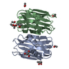
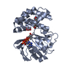
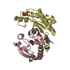
| Title | The Crystal structure of Sclerotium rolfsii lectin in complex with N-acetyl-D-glucosamine | ||||||
 Components Components | Sclerotium rolfsii lectin | ||||||
 Keywords Keywords | SUGAR BINDING PROTEIN /  Lectin / dual specificity Lectin / dual specificity | ||||||
| Function / homology | Cytolysin/lectin / Mutm (Fpg) Protein; Chain: A, domain 2 / Sandwich / Mainly Beta / ACETATE ION Function and homology information Function and homology information | ||||||
| Biological species |  Athelia rolfsii (fungus) Athelia rolfsii (fungus) | ||||||
| Method | X-RAY DIFFRACTION / SYNCHROTRON / FOURIER SYNTHESIS / Resolution: 1.7 Å | ||||||
 Authors Authors | Leonidas, D.D. / Zographos, S.E. / Oikonomakos, N.G. | ||||||
 Citation Citation | Journal: J.Mol.Biol. / Year: 2007 Title: Structural Basis for the Carbohydrate Recognition of the Sclerotium rolfsii Lectin Authors: Leonidas, D.D. / Swamy, B.M. / Hatzopoulos, G.N. / Gonchigar, S.J. / Chachadi, V.B. / Inamdar, S.R. / Zographos, S.E. / Oikonomakos, N.G. | ||||||
| History |
| ||||||
| Remark 999 | SEQUENCE AT THE TIME OF PROCESSING, THE SEQUENCE OF THIS PROTEIN IS NOT AVAILABLE AT THE UNP ...SEQUENCE AT THE TIME OF PROCESSING, THE SEQUENCE OF THIS PROTEIN IS NOT AVAILABLE AT THE UNP SEQUENCE DATABASE. |
- Structure visualization
| Structure viewer | Molecule: MolmilJmol/JSmol |
|---|
- Downloads & links
-Download
| PDBx/mmCIF format | 2ofe.cif.gz | 80.5 KB | Display | PDBx/mmCIF format |
|---|---|---|---|---|
| PDB format | pdb2ofe.ent.gz | 59.5 KB | Display | PDB format |
| PDBx/mmJSON format | 2ofe.json.gz | Tree view | PDBx/mmJSON format | |
| Others |  Other downloads Other downloads |
-Validation report
| Arichive directory | https://data.pdbj.org/pub/pdb/validation_reports/of/2ofeftp://data.pdbj.org/pub/pdb/validation_reports/of/2ofe | HTTPS FTP |
|---|
-Related structure data
| Related structure data |  2ofcSC 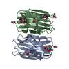 2ofdC S: Starting model for refinement C: citing same article ( |
|---|---|
| Similar structure data |


-Links
 PDBj
PDBj- Assembly
| Deposited unit | 
| ||||||||||||
|---|---|---|---|---|---|---|---|---|---|---|---|---|---|
| 1 |
| ||||||||||||
| 2 | 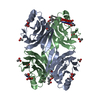
| ||||||||||||
| Unit cell |
| ||||||||||||
| Components on special symmetry positions |
| ||||||||||||
| Details | The Biological assembly of SRL is a dimer which is included in the asymmetric unit |
-Components
| #1: Protein | Mass: 16089.714 Da / Num. of mol.: 2 / Source method: isolated from a natural source / Source: (natural) Athelia rolfsii (fungus)#2: Sugar | N-Acetylglucosamine  Type: D-saccharide, beta linking / Mass: 221.208 Da / Num. of mol.: 2 Type: D-saccharide, beta linking / Mass: 221.208 Da / Num. of mol.: 2Source method: isolated from a genetically manipulated source Formula: C8H15NO6 #3: Chemical | Acetate  Mass: 59.044 Da / Num. of mol.: 3 / Source method: obtained synthetically / Formula: C2H3O2 Mass: 59.044 Da / Num. of mol.: 3 / Source method: obtained synthetically / Formula: C2H3O2#4: Chemical | 2-Methyl-2,4-pentanediol  Mass: 118.174 Da / Num. of mol.: 3 / Source method: obtained synthetically / Formula: C6H14O2 / Comment: precipitant*YM Mass: 118.174 Da / Num. of mol.: 3 / Source method: obtained synthetically / Formula: C6H14O2 / Comment: precipitant*YM#5: Water | ChemComp-HOH / | Water Mass: 18.015 Da / Num. of mol.: 379 / Source method: isolated from a natural source / Formula: H2O Mass: 18.015 Da / Num. of mol.: 379 / Source method: isolated from a natural source / Formula: H2O |
|---|
-Experimental details
-Experiment
| Experiment | Method: X-RAY DIFFRACTION / Number of used crystals: 1 |
|---|
- Sample preparation
| Crystal | Density Matthews: 2.48 Å3/Da / Density % sol: 50.35 % |
|---|---|
| Crystal grow | Temperature: 289 K / Method: vapor diffusion, hanging drop / pH: 8.5 Details: 0.2 M ammonium acetate, 30 % MPD, 0.1 M Tris/HCl, pH 8.5, VAPOR DIFFUSION, HANGING DROP, temperature 289K |
-Data collection
| Diffraction | Mean temperature: 100 K |
|---|---|
| Diffraction source | Source: SYNCHROTRON / Site: SRS  / Beamline: PX14.1 / Wavelength: 1.488 Å / Beamline: PX14.1 / Wavelength: 1.488 Å |
| Detector | Type: ADSC QUANTUM 4 / Detector: CCD / Date: Oct 9, 2006 |
| Radiation | Protocol: SINGLE WAVELENGTH / Monochromatic (M) / Laue (L): M / Scattering type: x-ray |
| Radiation wavelength | Wavelength: 1.488 Å / Relative weight: 1 |
| Reflection | Resolution: 1.7→30 Å / Num. all: 34881 / Num. obs: 34881 / % possible obs: 96.9 % / Observed criterion σ(F): 0 / Observed criterion σ(I): -3 / Redundancy: 5.5 % / Biso Wilson estimate: 8.994 Å2 / Rsym value: 0.055 / Net I/σ(I): 22.1 |
| Reflection shell | Resolution: 1.7→1.73 Å / Redundancy: 5.4 % / Mean I/σ(I) obs: 19 / Num. unique all: 1734 / Rsym value: 0.066 / % possible all: 98.2 |
- Processing
| Software |
| ||||||||||||||||||||||||||||||||||||||||||||||||||||||||||||||||||||||||||||||||||||||||||
|---|---|---|---|---|---|---|---|---|---|---|---|---|---|---|---|---|---|---|---|---|---|---|---|---|---|---|---|---|---|---|---|---|---|---|---|---|---|---|---|---|---|---|---|---|---|---|---|---|---|---|---|---|---|---|---|---|---|---|---|---|---|---|---|---|---|---|---|---|---|---|---|---|---|---|---|---|---|---|---|---|---|---|---|---|---|---|---|---|---|---|---|
| Refinement | Method to determine structure: FOURIER SYNTHESIS Starting model: PDB entry 2OFC Resolution: 1.7→30 Å / Cor.coef. Fo:Fc: 0.949 / Cor.coef. Fo:Fc free: 0.929 / SU B: 1.436 / SU ML: 0.049 / Cross valid method: THROUGHOUT / σ(F): 0 / ESU R: 0.097 / ESU R Free: 0.093 / Stereochemistry target values: MAXIMUM LIKELIHOOD / Details: HYDROGENS HAVE BEEN ADDED IN THE RIDING POSITIONS
| ||||||||||||||||||||||||||||||||||||||||||||||||||||||||||||||||||||||||||||||||||||||||||
| Solvent computation | Ion probe radii: 0.8 Å / Shrinkage radii: 0.8 Å / VDW probe radii: 1.4 Å / Solvent model: MASK | ||||||||||||||||||||||||||||||||||||||||||||||||||||||||||||||||||||||||||||||||||||||||||
| Displacement parameters | Biso mean: 9.121 Å2
| ||||||||||||||||||||||||||||||||||||||||||||||||||||||||||||||||||||||||||||||||||||||||||
| Refine analyze |
| ||||||||||||||||||||||||||||||||||||||||||||||||||||||||||||||||||||||||||||||||||||||||||
| Refinement step | Cycle: LAST / Resolution: 1.7→30 Å
| ||||||||||||||||||||||||||||||||||||||||||||||||||||||||||||||||||||||||||||||||||||||||||
| Refine LS restraints |
| ||||||||||||||||||||||||||||||||||||||||||||||||||||||||||||||||||||||||||||||||||||||||||
| LS refinement shell | Resolution: 1.7→1.747 Å / Total num. of bins used: 20
|
