- PDB-4yjw: Crystal structure of a DUF3829 family protein (BVU_3067) from Bac... -
+
Open data
ID or keywords:
Loading...
-
Basic information
Entry
Database: PDB / ID: 4yjw
Title
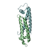
Crystal structure of a DUF3829 family protein (BVU_3067) from Bacteroides vulgatus ATCC 8482 at 1.80 A resolution
Components
DUF3829 family protein
Keywords
STRUCTURAL GENOMICS UNKNOWN FUNCTION / All alpha protein / PF12889 family / Structural Genomics / Joint Center for Structural Genomics / JCSG / Protein Structure Initiative / PSI-BIOLOGY / UNKNOWN FUNCTION
Function / homology
Uncharacterised protein PF12889, N-terminal DUF3829 / Four Helix Bundle (Hemerythrin (Met), subunit A) / Up-down Bundle / Mainly Alpha / Uncharacterized protein
Function and homology information
Biological species
Bacteroides vulgatus (bacteria)
Method
X-RAY DIFFRACTION / SYNCHROTRON / SAD / Resolution: 1.8 Å
Monochromator: double crystal Si(111) / Protocol: SINGLE WAVELENGTH / Monochromatic (M) / Laue (L): M / Scattering type: x-ray
Radiation wavelength
Wavelength: 0.9792 Å / Relative weight: 1
Reflection
Resolution: 1.8→28.652 Å / Num. obs: 16483 / % possible obs: 100 % / Observed criterion σ(I): -3 / Redundancy: 6.17 % / Biso Wilson estimate: 21.665 Å2 / Rmerge F obs: 0.999 / Rmerge(I) obs: 0.08 / Rrim(I) all: 0.088 / Net I/σ(I): 16.13 / Num. measured all: 187648
Reflection shell
Resolution (Å)
Rmerge F obs
Rmerge(I) obs
Mean I/σ(I) obs
Num. measured obs
Num. possible
Num. unique obs
Rrim(I) all
Diffraction-ID
% possible all
1.8-1.86
0.651
0.739
1.9
12263
2859
2858
0.843
1
100
1.86-1.94
0.817
0.605
2.9
19008
3223
3223
0.664
100
1.94-2.03
0.909
0.427
4.4
19963
3117
3117
0.465
100
2.03-2.13
0.961
0.268
6.9
18244
2843
2843
0.291
100
2.13-2.27
0.983
0.182
10
20532
3194
3194
0.199
100
2.27-2.44
0.991
0.139
12.8
19050
2952
2952
0.151
100
2.44-2.69
0.994
0.105
16.7
19929
3085
3085
0.114
100
2.69-3.07
0.997
0.074
22.6
19338
2992
2992
0.08
100
3.07-3.87
0.999
0.043
35.6
19617
3057
3057
0.047
100
3.87-28.652
0.999
0.031
47
19704
3084
3073
0.034
99.6
-
Phasing
Phasing
Method: SAD
-
Processing
Software
Name
Version
Classification
PDB_EXTRACT
3.1
dataextraction
SHELX
phasing
SHARP
phasing
XDS
November3, 2014BUILT=20141118
datascaling
BUSTER-TNT
2.10.2
refinement
XSCALE
datascaling
SHELXD
phasing
BUSTER
2.10.2
refinement
Refinement
Method to determine structure: SAD / Resolution: 1.8→28.652 Å / Cor.coef. Fo:Fc: 0.9532 / Cor.coef. Fo:Fc free: 0.9459 / Occupancy max: 1 / Occupancy min: 0.3 / Cross valid method: THROUGHOUT / σ(F): 0 Details: 1. A MET-INHIBITION PROTOCOL WAS USED FOR SELENOMETHIONINE INCORPORATION DURING PROTEIN EXPRESSION. THE OCCUPANCY OF THE SE ATOMS IN THE MSE RESIDUES WAS REDUCED TO 0.75 FOR THE REDUCED ...Details: 1. A MET-INHIBITION PROTOCOL WAS USED FOR SELENOMETHIONINE INCORPORATION DURING PROTEIN EXPRESSION. THE OCCUPANCY OF THE SE ATOMS IN THE MSE RESIDUES WAS REDUCED TO 0.75 FOR THE REDUCED SCATTERING POWER DUE TO PARTIAL S-MET INCORPORATION. 2. THE SAD PHASES WERE USED AS RESTRAINTS DURING REFINEMENT. 3. A UNMODELED DENSITY BLOB AT THE TWO-FOLD AXIS (NEAR A62) COULD BE EXPLAINED BY A BENZOIC ACID-LIKE COMPOUND.
In the structure databanks used in Yorodumi, some data are registered as the other names, "COVID-19 virus" and "2019-nCoV". Here are the details of the virus and the list of structure data.
Jan 31, 2019. EMDB accession codes are about to change! (news from PDBe EMDB page)
EMDB accession codes are about to change! (news from PDBe EMDB page)
The allocation of 4 digits for EMDB accession codes will soon come to an end. Whilst these codes will remain in use, new EMDB accession codes will include an additional digit and will expand incrementally as the available range of codes is exhausted. The current 4-digit format prefixed with “EMD-” (i.e. EMD-XXXX) will advance to a 5-digit format (i.e. EMD-XXXXX), and so on. It is currently estimated that the 4-digit codes will be depleted around Spring 2019, at which point the 5-digit format will come into force.
The EM Navigator/Yorodumi systems omit the EMD- prefix.
Related info.:Q: What is EMD? / ID/Accession-code notation in Yorodumi/EM Navigator
Yorodumi is a browser for structure data from EMDB, PDB, SASBDB, etc.
This page is also the successor to EM Navigator detail page, and also detail information page/front-end page for Omokage search.
The word "yorodu" (or yorozu) is an old Japanese word meaning "ten thousand". "mi" (miru) is to see.
Related info.:EMDB / PDB / SASBDB / Comparison of 3 databanks / Yorodumi Search / Aug 31, 2016. New EM Navigator & Yorodumi / Yorodumi Papers / Jmol/JSmol / Function and homology information / Changes in new EM Navigator and Yorodumi
 Movie
Movie Controller

 Yorodumi
 Open data
 Basic information
 Components
 Keywords
 Structural Genomics / Joint Center for Structural Genomics / JCSG /
  Function and homology information
 Bacteroides vulgatus (bacteria)
 Authors
 Citation
 Structure visualization
 Downloads & links
 Other downloads
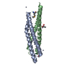


 PDBj
PDBj Assembly
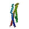
 Mass: 18.015 Da / Num. of mol.: 184 / Source method: isolated from a natural source / Formula: H2O
 Sample preparation
 / Beamline: BL14-1 / Wavelength: 0.9792 Å
 Processing