 Movie
Movie Controller

[English] 日本語
 Yorodumi
- PDB-4pac: Crystal Structure of Histidine-containing Phosphotransfer Protein... -
+ Open data

- Basic information
| Entry | Database: PDB / ID: 4pac | |||||||||||||||||||||
|---|---|---|---|---|---|---|---|---|---|---|---|---|---|---|---|---|---|---|---|---|---|---|
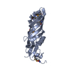
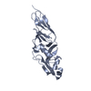
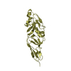
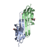
| Title | Crystal Structure of Histidine-containing Phosphotransfer Protein AHP2 from Arabidopsis thaliana | |||||||||||||||||||||
 Components Components | Histidine-containing phosphotransfer protein 2 | |||||||||||||||||||||
 Keywords Keywords |  SIGNALING PROTEIN / cytokinin signaling / helical fold SIGNALING PROTEIN / cytokinin signaling / helical fold | |||||||||||||||||||||
| Function / homology |  Function and homology information Function and homology informationantipodal cell differentiation / embryo sac egg cell differentiation / protein histidine kinase binding / cytokinin-activated signaling pathway / histidine phosphotransfer kinase activity / phosphorelay signal transduction system / phosphorylation / nucleus / cytosol / cytoplasmSimilarity search - Function | |||||||||||||||||||||
| Biological species |  Arabidopsis thaliana (thale cress) Arabidopsis thaliana (thale cress) | |||||||||||||||||||||
| Method | X-RAY DIFFRACTION / SYNCHROTRON / SIRAS / Resolution: 2.53 Å | |||||||||||||||||||||
 Authors Authors | Degtjarik, O. / Dopitova, R. / Puehringer, S. / Weiss, M.S. / Janda, L. / Hejatko, J. / Kuta-Smatanova, I. | |||||||||||||||||||||
| Funding support |  Czech Republic, 6items Czech Republic, 6items
| |||||||||||||||||||||
 Citation Citation | Journal: To Be Published Title: Crystal structure of AHP2 from Arabidopsis thaliana Authors: Degtjarik, O. / Dopitova, R. / Puehringer, S. / Reha, D. / Otrusinova, O. / Pekarova, B. / Kuty, M. / Zidek, L. / Janda, L. / Weiss, M.S. / Kuta-Smatanova, I. / Hejatko, H. | |||||||||||||||||||||
| History |
|
- Structure visualization
| Structure viewer | Molecule: MolmilJmol/JSmol |
|---|
- Downloads & links
-Download
| PDBx/mmCIF format | 4pac.cif.gz | 45.7 KB | Display | PDBx/mmCIF format |
|---|---|---|---|---|
| PDB format | pdb4pac.ent.gz | 31.3 KB | Display | PDB format |
| PDBx/mmJSON format | 4pac.json.gz | Tree view | PDBx/mmJSON format | |
| Others |  Other downloads Other downloads |
-Validation report
| Arichive directory | https://data.pdbj.org/pub/pdb/validation_reports/pa/4pacftp://data.pdbj.org/pub/pdb/validation_reports/pa/4pac | HTTPS FTP |
|---|
-Related structure data
| Related structure data | |
|---|---|
| Similar structure data |


-Links
 PDBj
PDBj


- Assembly
| Deposited unit | 
| ||||||||
|---|---|---|---|---|---|---|---|---|---|
| 1 |
| ||||||||
| Unit cell |
|
-Components
| #1: Protein | Mass: 22369.232 Da / Num. of mol.: 1 Source method: isolated from a genetically manipulated source Source: (gene. exp.) Arabidopsis thaliana (thale cress) / Gene: AHP2, ATHP1, At3g29350, MUO10.6 / Plasmid: pRSET B / Production host:  Escherichia coli (E. coli) / Strain (production host): BL21(DE3) / Variant (production host): pLysS / References: UniProt: Q9ZNV8 Escherichia coli (E. coli) / Strain (production host): BL21(DE3) / Variant (production host): pLysS / References: UniProt: Q9ZNV8 |
|---|---|
| #2: Chemical | ChemComp-MES / MES (buffer)  Mass: 195.237 Da / Num. of mol.: 1 / Source method: obtained synthetically / Formula: C6H13NO4S / Comment: pH buffer*YM Mass: 195.237 Da / Num. of mol.: 1 / Source method: obtained synthetically / Formula: C6H13NO4S / Comment: pH buffer*YM |
| #3: Chemical | ChemComp-IMD / Imidazole  Mass: 69.085 Da / Num. of mol.: 1 / Source method: obtained synthetically / Formula: C3H5N2 Mass: 69.085 Da / Num. of mol.: 1 / Source method: obtained synthetically / Formula: C3H5N2 |
| #4: Water | ChemComp-HOH / Water Mass: 18.015 Da / Num. of mol.: 18 / Source method: isolated from a natural source / Formula: H2O Mass: 18.015 Da / Num. of mol.: 18 / Source method: isolated from a natural source / Formula: H2O |
-Experimental details
-Experiment
| Experiment | Method: X-RAY DIFFRACTION / Number of used crystals: 1 |
|---|
- Sample preparation
| Crystal | Density Matthews: 3.4 Å3/Da / Density % sol: 63.84 % |
|---|---|
| Crystal grow | Temperature: 277 K / Method: vapor diffusion / pH: 6.5 / Details: 0.1M MES, 1.6M magnesium sulfate / PH range: 5.8 - 6.5 |
-Data collection
| Diffraction | Mean temperature: 100 K |
|---|---|
| Diffraction source | Source: SYNCHROTRON / Site: BESSY  / Beamline: 14.2 / Wavelength: 0.91841 Å / Beamline: 14.2 / Wavelength: 0.91841 Å |
| Detector | Type: RAYONIX MX-225 / Detector: CCD / Date: Feb 28, 2012 |
| Radiation | Protocol: SINGLE WAVELENGTH / Monochromatic (M) / Laue (L): M / Scattering type: x-ray |
| Radiation wavelength | Wavelength: 0.91841 Å / Relative weight: 1 |
| Reflection | Resolution: 2.53→50 Å / Num. obs: 10808 / % possible obs: 99.6 % / Redundancy: 7 % / Net I/σ(I): 15.7 |
| Reflection shell | Resolution: 2.53→2.68 Å / % possible obs: 98.7 % / Redundancy: 7.02 % / Rmerge(I) obs: 1.014 / Mean I/σ(I) obs: 1.76 |
- Processing
| Software |
| ||||||||||||||||||||||||||||||||||||||||||||||||||||||||||||||||||||||||||||||||||||||||||||||||||||||||||||||||||||||||||||||||||||||||||||||||||||||||||||||||||||||||||||||||||||||
|---|---|---|---|---|---|---|---|---|---|---|---|---|---|---|---|---|---|---|---|---|---|---|---|---|---|---|---|---|---|---|---|---|---|---|---|---|---|---|---|---|---|---|---|---|---|---|---|---|---|---|---|---|---|---|---|---|---|---|---|---|---|---|---|---|---|---|---|---|---|---|---|---|---|---|---|---|---|---|---|---|---|---|---|---|---|---|---|---|---|---|---|---|---|---|---|---|---|---|---|---|---|---|---|---|---|---|---|---|---|---|---|---|---|---|---|---|---|---|---|---|---|---|---|---|---|---|---|---|---|---|---|---|---|---|---|---|---|---|---|---|---|---|---|---|---|---|---|---|---|---|---|---|---|---|---|---|---|---|---|---|---|---|---|---|---|---|---|---|---|---|---|---|---|---|---|---|---|---|---|---|---|---|---|
| Refinement | Method to determine structure: SIRAS / Resolution: 2.53→50 Å / Cor.coef. Fo:Fc: 0.941 / Cor.coef. Fo:Fc free: 0.914 / SU B: 11.856 / SU ML: 0.242 / Cross valid method: THROUGHOUT / ESU R: 0.353 / ESU R Free: 0.31 / Stereochemistry target values: MAXIMUM LIKELIHOOD / Details: HYDROGENS HAVE BEEN USED IF PRESENT IN THE INPUT
| ||||||||||||||||||||||||||||||||||||||||||||||||||||||||||||||||||||||||||||||||||||||||||||||||||||||||||||||||||||||||||||||||||||||||||||||||||||||||||||||||||||||||||||||||||||||
| Solvent computation | Ion probe radii: 0.8 Å / Shrinkage radii: 0.8 Å / VDW probe radii: 1.2 Å / Solvent model: MASK | ||||||||||||||||||||||||||||||||||||||||||||||||||||||||||||||||||||||||||||||||||||||||||||||||||||||||||||||||||||||||||||||||||||||||||||||||||||||||||||||||||||||||||||||||||||||
| Displacement parameters | Biso mean: 59.628 Å2
| ||||||||||||||||||||||||||||||||||||||||||||||||||||||||||||||||||||||||||||||||||||||||||||||||||||||||||||||||||||||||||||||||||||||||||||||||||||||||||||||||||||||||||||||||||||||
| Refinement step | Cycle: 1 / Resolution: 2.53→50 Å
| ||||||||||||||||||||||||||||||||||||||||||||||||||||||||||||||||||||||||||||||||||||||||||||||||||||||||||||||||||||||||||||||||||||||||||||||||||||||||||||||||||||||||||||||||||||||
| Refine LS restraints |
|