 Movie
Movie Controller

[English] 日本語
 Yorodumi
- PDB-4g78: Subatomic Resolution Crystal Structure of Histidine-containing Ph... -
+ Open data

- Basic information
| Entry | Database: PDB / ID: 4g78 | ||||||
|---|---|---|---|---|---|---|---|
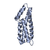
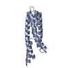
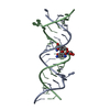
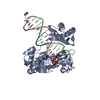
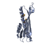
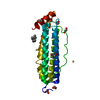
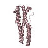
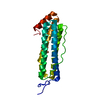
| Title | Subatomic Resolution Crystal Structure of Histidine-containing Phosphotransfer Protein MtHPt2 from Medicago truncatula | ||||||
 Components Components | Histidine phosphotransfer protein | ||||||
 Keywords Keywords |  TRANSFERASE / SUBATOMIC RESOLUTION / HELIX BUNDLE / PLANT HORMONE SIGNAL TRANSDUCTION / HISTIDINE KINASE TRANSFERASE / SUBATOMIC RESOLUTION / HELIX BUNDLE / PLANT HORMONE SIGNAL TRANSDUCTION / HISTIDINE KINASE | ||||||
| Function / homology |  Function and homology information Function and homology informationprotein histidine kinase binding / cytokinin-activated signaling pathway / histidine phosphotransfer kinase activity / phosphorelay signal transduction system / phosphorylation / nucleus / cytosol / cytoplasmSimilarity search - Function | ||||||
| Biological species |  Medicago truncatula (barrel medic) Medicago truncatula (barrel medic) | ||||||
| Method | X-RAY DIFFRACTION / SYNCHROTRON / MOLECULAR REPLACEMENT / molecular replacement / Resolution: 0.92 Å | ||||||
 Authors Authors | Ruszkowski, M. / Sikorski, M. / Jaskolski, M. | ||||||
 Citation Citation | Journal: To be Published Title: Subatomic Resolution Crystal Structure of Histidine-containing Phosphotransfer Protein MtHPt2 from Medicago truncatula Authors: Ruszkowski, M. / Sikorski, M. / Jaskolski, M. #1: Journal: Plant Cell / Year: 2006 Title: The Arabidopsis histidine phosphotransfer proteins are redundant positive regulators of cytokinin signaling. Authors: Hutchison, C.E. / Li, J. / Argueso, C. / Gonzalez, M. / Lee, E. / Lewis, M.W. / Maxwell, B.B. / Perdue, T.D. / Schaller, G.E. / Alonso, J.M. / Ecker, J.R. / Kieber, J.J. #2: Journal: Protein Sci. / Year: 2005Title: Crystal structure of the histidine-containing phosphotransfer protein ZmHP2 from maize. Authors: Sugawara, H. / Kawano, Y. / Hatakeyama, T. / Yamaya, T. / Kamiya, N. / Sakakibara, H. #3: Journal: J.Mol.Biol. / Year: 1999Title: Conservation of structure and function among histidine-containing phosphotransfer (HPt) domains as revealed by the crystal structure of YPD1. Authors: Xu, Q. / West, A.H. | ||||||
| History |
|
- Structure visualization
| Structure viewer | Molecule: MolmilJmol/JSmol |
|---|
- Downloads & links
-Download
| PDBx/mmCIF format | 4g78.cif.gz | 88.8 KB | Display | PDBx/mmCIF format |
|---|---|---|---|---|
| PDB format | pdb4g78.ent.gz | 67.5 KB | Display | PDB format |
| PDBx/mmJSON format | 4g78.json.gz | Tree view | PDBx/mmJSON format | |
| Others |  Other downloads Other downloads |
-Validation report
| Arichive directory | https://data.pdbj.org/pub/pdb/validation_reports/g7/4g78ftp://data.pdbj.org/pub/pdb/validation_reports/g7/4g78 | HTTPS FTP |
|---|
-Related structure data
| Related structure data |  3us6S S: Starting model for refinement |
|---|---|
| Similar structure data |


-Links
 PDBj
PDBj- Assembly
| Deposited unit | 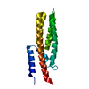
| ||||||||
|---|---|---|---|---|---|---|---|---|---|
| 1 |
| ||||||||
| Unit cell |
|
-Components
| #1: Protein | Mass: 17475.738 Da / Num. of mol.: 1 Source method: isolated from a genetically manipulated source Source: (gene. exp.) Medicago truncatula (barrel medic) / Gene: MtHPt2, MTR_1g089130, MTR_1g089350 / Plasmid: pMCSG48 / Production host:  Escherichia coli (E. coli) / Strain (production host): BL21 Magic Escherichia coli (E. coli) / Strain (production host): BL21 MagicReferences: UniProt: G7I2T8, Transferases; Transferring phosphorus-containing groups; Phosphotransferases with a nitrogenous group as acceptor |
|---|---|
| #2: Water | ChemComp-HOH / Water Mass: 18.015 Da / Num. of mol.: 207 / Source method: isolated from a natural source / Formula: H2O Mass: 18.015 Da / Num. of mol.: 207 / Source method: isolated from a natural source / Formula: H2O |
-Experimental details
-Experiment
| Experiment | Method: X-RAY DIFFRACTION / Number of used crystals: 1 |
|---|
- Sample preparation
| Crystal | Density Matthews: 1.81 Å3/Da / Density % sol: 32.01 % |
|---|---|
| Crystal grow | Temperature: 292 K / pH: 6.5 Details: 0.09M Halogens, 0.1M buffer system 1, 30% PEG 550MME + PEG 20K (Molecular Dimensions Morpheus Screen B1), pH 6.5, VAPOR DIFFUSION, HANGING DROP, temperature 292K |
-Data collection
| Diffraction | Mean temperature: 100 K |
|---|---|
| Diffraction source | Source: SYNCHROTRON / Site: BESSY  / Beamline: 14.2 / Wavelength: 0.91841 / Beamline: 14.2 / Wavelength: 0.91841 |
| Detector | Type: MARMOSAIC 225 mm CCD / Detector: CCD / Date: Mar 17, 2012 / Details: FOCUSING MIRRORS |
| Radiation | Monochromator: SAGITALLY FOCUSED SI(111) / Protocol: SINGLE WAVELENGTH / Monochromatic (M) / Laue (L): M / Scattering type: x-ray |
| Radiation wavelength | Wavelength: 0.91841 Å / Relative weight: 1 |
| Reflection | Resolution: 0.92→50 Å / Num. obs: 84634 / % possible obs: 97.8 % / Observed criterion σ(I): -3 / Redundancy: 6.3 % / Biso Wilson estimate: 13.232 Å2 / Rmerge(I) obs: 0.036 / Net I/σ(I): 21.78 |
| Reflection shell | Resolution: 0.92→0.94 Å / Rmerge(I) obs: 0.738 / Mean I/σ(I) obs: 1.89 / % possible all: 75.1 |
-Phasing
| Phasing | Method: molecular replacement | |||||||||
|---|---|---|---|---|---|---|---|---|---|---|
| Phasing MR |
|
- Processing
| Software |
| |||||||||||||||||||||||||||||||||
|---|---|---|---|---|---|---|---|---|---|---|---|---|---|---|---|---|---|---|---|---|---|---|---|---|---|---|---|---|---|---|---|---|---|---|
| Refinement | Method to determine structure: MOLECULAR REPLACEMENT Starting model: PDB ENTRY 3US6 Resolution: 0.92→50 Å / Cross valid method: R-FREE / Stereochemistry target values: Engh & Huber / Details: HYDROGEN ATOMS WERE ADDED AT RIDING POSITIONS
| |||||||||||||||||||||||||||||||||
| Solvent computation | Solvent model: BABINET MODEL | |||||||||||||||||||||||||||||||||
| Displacement parameters | Biso mean: 19.627 Å2 | |||||||||||||||||||||||||||||||||
| Refinement step | Cycle: LAST / Resolution: 0.92→50 Å
| |||||||||||||||||||||||||||||||||
| Refine LS restraints |
| |||||||||||||||||||||||||||||||||
| LS refinement shell | Resolution: 0.92→0.96 Å / Rfactor Rwork: 0.271 |
