 Movie
Movie Controller


+ Open data

- Basic information
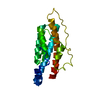
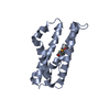
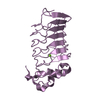
| Entry | Database: PDB / ID: 1qsp | ||||||
|---|---|---|---|---|---|---|---|
| Title | CRYSTAL STRUCTURE OF THE YEAST PHOSPHORELAY PROTEIN YPD1 | ||||||
 Components Components | YPD1 | ||||||
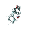
 Keywords Keywords |  SIGNALING PROTEIN / FOUR-HELIX BUNDLE / HISTIDINE-CONTAINING PHOSPHOTRANSFER (HPT) DOMAIN / TWO- COMPONENT SIGNAL TRANSDUCTION SIGNALING PROTEIN / FOUR-HELIX BUNDLE / HISTIDINE-CONTAINING PHOSPHOTRANSFER (HPT) DOMAIN / TWO- COMPONENT SIGNAL TRANSDUCTION | ||||||
| Function / homology |  Function and homology information Function and homology informationprotein histidine kinase binding / transferase activity, transferring phosphorus-containing groups / histidine phosphotransfer kinase activity / osmosensory signaling via phosphorelay pathway / phosphorelay signal transduction system / phosphorylation / nucleus / cytoplasmSimilarity search - Function | ||||||
| Biological species |  Saccharomyces cerevisiae (brewer's yeast) Saccharomyces cerevisiae (brewer's yeast) | ||||||
| Method | X-RAY DIFFRACTION / Resolution: 2.7 Å | ||||||
 Authors Authors | Xu, Q. / West, A.H. | ||||||
 Citation Citation | Journal: J.Mol.Biol. / Year: 1999 Title: Conservation of structure and function among histidine-containing phosphotransfer (HPt) domains as revealed by the crystal structure of YPD1. Authors: Xu, Q. / West, A.H. #1: Journal: Acta Crystallogr.,Sect.D / Year: 1999Title: Purification, Crystallization and Preliminary X-ray Diffraction Analysis of the Yeast Phosphorelay Protein YPD1 Authors: Xu, Q. / West, A.H. | ||||||
| History |
|
- Structure visualization
| Structure viewer | Molecule: MolmilJmol/JSmol |
|---|
- Downloads & links
-Download
| PDBx/mmCIF format | 1qsp.cif.gz | 77 KB | Display | PDBx/mmCIF format |
|---|---|---|---|---|
| PDB format | pdb1qsp.ent.gz | 59.8 KB | Display | PDB format |
| PDBx/mmJSON format | 1qsp.json.gz | Tree view | PDBx/mmJSON format | |
| Others |  Other downloads Other downloads |
-Validation report
| Arichive directory | https://data.pdbj.org/pub/pdb/validation_reports/qs/1qspftp://data.pdbj.org/pub/pdb/validation_reports/qs/1qsp | HTTPS FTP |
|---|
-Related structure data
| Similar structure data |
|---|


-Links
 PDBj
PDBj- Assembly
| Deposited unit | 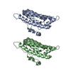
| ||||||||
|---|---|---|---|---|---|---|---|---|---|
| 1 | 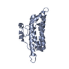
| ||||||||
| 2 | 
| ||||||||
| Unit cell |
|
-Components
| #1: Protein | Mass: 18969.379 Da / Num. of mol.: 2 Source method: isolated from a genetically manipulated source Source: (gene. exp.) Saccharomyces cerevisiae (brewer's yeast)Production host:  Escherichia coli (E. coli) / References: UniProt: Q07688 Escherichia coli (E. coli) / References: UniProt: Q07688#2: Water | ChemComp-HOH / | Water Mass: 18.015 Da / Num. of mol.: 104 / Source method: isolated from a natural source / Formula: H2O Mass: 18.015 Da / Num. of mol.: 104 / Source method: isolated from a natural source / Formula: H2O |
|---|
-Experimental details
-Experiment
| Experiment | Method: X-RAY DIFFRACTION / Number of used crystals: 1 |
|---|
- Sample preparation
| Crystal | Density Matthews: 2.23 Å3/Da / Density % sol: 44.93 % | |||||||||||||||||||||||||
|---|---|---|---|---|---|---|---|---|---|---|---|---|---|---|---|---|---|---|---|---|---|---|---|---|---|---|
| Crystal grow | Temperature: 298 K / Method: vapor diffusion, hanging drop / pH: 5 Details: PEG 4000, SODIUM ACETATE AMMONIUM ACETATE, BETA-MERCAPTOETHANOL, pH 5.0, VAPOR DIFFUSION, HANGING DROP, temperature 298K | |||||||||||||||||||||||||
| Crystal grow | *PLUS Details: Xu, Q., (1999) Acta Crystallogr., Sect.D, 55, 291. | |||||||||||||||||||||||||
| Components of the solutions | *PLUS
|
-Data collection
| Diffraction | Mean temperature: 298 K |
|---|---|
| Diffraction source | Source: ROTATING ANODE / Type: RIGAKU RU200 / Wavelength: 1.5418 |
| Detector | Type: RIGAKU RAXIS II / Detector: IMAGE PLATE / Date: Nov 20, 1997 |
| Radiation | Protocol: SINGLE WAVELENGTH / Monochromatic (M) / Laue (L): M / Scattering type: x-ray |
| Radiation wavelength | Wavelength: 1.5418 Å / Relative weight: 1 |
| Reflection | Resolution: 2.7→30 Å / Num. all: 9674 / Num. obs: 52152 / % possible obs: 91.6 % / Observed criterion σ(F): 0 / Observed criterion σ(I): 0 / Redundancy: 5.4 % / Biso Wilson estimate: 27.752 Å2 / Rmerge(I) obs: 0.066 / Net I/σ(I): 9.7 |
| Reflection shell | Resolution: 2.7→2.85 Å / Redundancy: 2.9 % / Rmerge(I) obs: 0.078 / % possible all: 82 |
| Reflection | *PLUS Num. all: 52152 / Num. obs: 9674 |
| Reflection shell | *PLUS % possible obs: 82 % / Mean I/σ(I) obs: 11.2 |
- Processing
| Software |
| ||||||||||||||||||||||||||||||||||||||||||||||||||||||||||||
|---|---|---|---|---|---|---|---|---|---|---|---|---|---|---|---|---|---|---|---|---|---|---|---|---|---|---|---|---|---|---|---|---|---|---|---|---|---|---|---|---|---|---|---|---|---|---|---|---|---|---|---|---|---|---|---|---|---|---|---|---|---|
| Refinement | Resolution: 2.7→30 Å / σ(F): 0 / σ(I): 0 / Stereochemistry target values: ENGH & HUBER
| ||||||||||||||||||||||||||||||||||||||||||||||||||||||||||||
| Refinement step | Cycle: LAST / Resolution: 2.7→30 Å
| ||||||||||||||||||||||||||||||||||||||||||||||||||||||||||||
| Refine LS restraints |
| ||||||||||||||||||||||||||||||||||||||||||||||||||||||||||||
| Software | *PLUS Name: X-PLOR / Classification: refinement | ||||||||||||||||||||||||||||||||||||||||||||||||||||||||||||
| Refinement | *PLUS Highest resolution: 2.7 Å / Lowest resolution: 30 Å / σ(F): 0 | ||||||||||||||||||||||||||||||||||||||||||||||||||||||||||||
| Solvent computation | *PLUS | ||||||||||||||||||||||||||||||||||||||||||||||||||||||||||||
| Displacement parameters | *PLUS Biso mean: 27.681 Å2 | ||||||||||||||||||||||||||||||||||||||||||||||||||||||||||||
| Refine LS restraints | *PLUS Type: x_bond_d / Dev ideal: 0.007 |
