 Movie
Movie Controller

[English] 日本語
 Yorodumi
- PDB-3g9r: Structure of the HIV-1 gp41 Membrane-Proximal Ectodomain Region i... -
+ Open data

- Basic information
| Entry | Database: PDB / ID: 3g9r | ||||||
|---|---|---|---|---|---|---|---|
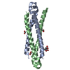
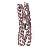
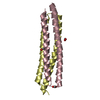
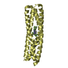
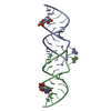
| Title | Structure of the HIV-1 gp41 Membrane-Proximal Ectodomain Region in a Putative Prefusion Conformation | ||||||
 Components Components | Fusion complex of HIV-1 Envelope glycoprotein and Saccharomyces cerevisiae General control protein GCN4 | ||||||
 Keywords Keywords |  VIRAL PROTEIN / gp41 / MPER / HIV-1 / membrane fusion / AIDS / Apoptosis / Cell membrane / Coiled coil / Envelope protein / Fusion protein / Host-virus interaction VIRAL PROTEIN / gp41 / MPER / HIV-1 / membrane fusion / AIDS / Apoptosis / Cell membrane / Coiled coil / Envelope protein / Fusion protein / Host-virus interaction | ||||||
| Function / homology |  Function and homology information Function and homology informationvirus-mediated perturbation of host defense response => GO:0019049 / : / protein localization to nuclear periphery / FCERI mediated MAPK activation / Activation of the AP-1 family of transcription factors / response to amino acid starvation / mediator complex binding / negative regulation of ribosomal protein gene transcription by RNA polymerase II / positive regulation of cellular response to amino acid starvation / Oxidative Stress Induced Senescence ...virus-mediated perturbation of host defense response => GO:0019049 / : / protein localization to nuclear periphery / FCERI mediated MAPK activation / Activation of the AP-1 family of transcription factors / response to amino acid starvation / mediator complex binding / negative regulation of ribosomal protein gene transcription by RNA polymerase II / positive regulation of cellular response to amino acid starvation / Oxidative Stress Induced Senescence / nitrogen catabolite activation of transcription from RNA polymerase II promoter / Dectin-2 family / TFIID-class transcription factor complex binding / positive regulation of transcription initiation by RNA polymerase II / amino acid biosynthetic process / positive regulation of RNA polymerase II transcription preinitiation complex assembly / positive regulation of plasma membrane raft polarization / positive regulation of receptor clustering / positive regulation of establishment of T cell polarity / cellular response to amino acid starvation / host cell endosome membrane / RNA polymerase II transcription regulator complex / : / DNA-binding transcription activator activity, RNA polymerase II-specific / clathrin-dependent endocytosis of virus by host cell / RNA polymerase II-specific DNA-binding transcription factor binding / transcription regulator complex / sequence-specific DNA binding / membrane => GO:0016020 / viral protein processing / DNA-binding transcription factor activity, RNA polymerase II-specific / regulation of cell cycle / intracellular signal transduction / RNA polymerase II cis-regulatory region sequence-specific DNA binding / DNA-binding transcription factor activity / fusion of virus membrane with host plasma membrane / virus-mediated perturbation of host defense response / fusion of virus membrane with host endosome membrane / viral envelope / chromatin binding / virion attachment to host cell / host cell plasma membrane / virion membrane / structural molecule activity / negative regulation of transcription by RNA polymerase II / positive regulation of transcription by RNA polymerase II / membrane / identical protein binding / nucleus / plasma membraneSimilarity search - Function | ||||||
| Biological species |   Human immunodeficiency virus 1 Human immunodeficiency virus 1 Saccharomyces cerevisiae (brewer's yeast) Saccharomyces cerevisiae (brewer's yeast) | ||||||
| Method | X-RAY DIFFRACTION / SYNCHROTRON / MOLECULAR REPLACEMENT / Resolution: 2 Å | ||||||
 Authors Authors | Liu, J. / Lu, M. | ||||||
 Citation Citation | Journal: Biochemistry / Year: 2009 Title: Structure of the HIV-1 gp41 Membrane-Proximal Ectodomain Region in a Putative Prefusion Conformation. Authors: Liu, J. / Deng, Y. / Dey, A. / Moore, J. / Lu, M. | ||||||
| History |
|
- Structure visualization
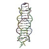
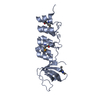
| Structure viewer | Molecule: MolmilJmol/JSmol |
|---|
- Downloads & links
-Download
| PDBx/mmCIF format | 3g9r.cif.gz | 126.1 KB | Display | PDBx/mmCIF format |
|---|---|---|---|---|
| PDB format | pdb3g9r.ent.gz | 101.9 KB | Display | PDB format |
| PDBx/mmJSON format | 3g9r.json.gz | Tree view | PDBx/mmJSON format | |
| Others |  Other downloads Other downloads |
-Validation report
| Arichive directory | https://data.pdbj.org/pub/pdb/validation_reports/g9/3g9rftp://data.pdbj.org/pub/pdb/validation_reports/g9/3g9r | HTTPS FTP |
|---|
-Related structure data
| Related structure data | 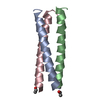 1gcmS S: Starting model for refinement |
|---|---|
| Similar structure data |

-Links
 PDBj
PDBj


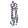
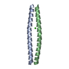
- Assembly
| Deposited unit | 
| ||||||||
|---|---|---|---|---|---|---|---|---|---|
| 1 | 
| ||||||||
| 2 | 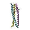
| ||||||||
| Unit cell |
|
-Components
| #1: Protein/peptide | Mass: 5415.377 Da / Num. of mol.: 6 Fragment: C-terminal MPER of HIV-1 GP41, Leucine-zipper domain of GCN4 Mutation: L27K, N30I, Y31K, H32R, L33I, V37I, A38K, L40I Source method: isolated from a genetically manipulated source Source: (gene. exp.) Human immunodeficiency virus 1, (gene. exp.) Saccharomyces cerevisiae (brewer's yeast)Production host:  Escherichia coli (E. coli) Escherichia coli (E. coli)References: UniProt: Q4QX39, UniProt: P03069, UniProt: P03377*PLUS #2: Chemical | ChemComp-PO4 / Phosphate  Mass: 94.971 Da / Num. of mol.: 5 / Source method: obtained synthetically / Formula: PO4 Mass: 94.971 Da / Num. of mol.: 5 / Source method: obtained synthetically / Formula: PO4#3: Chemical | ChemComp-MPD / ( 2-Methyl-2,4-pentanediol  Mass: 118.174 Da / Num. of mol.: 8 / Source method: obtained synthetically / Formula: C6H14O2 / Comment: precipitant*YM Mass: 118.174 Da / Num. of mol.: 8 / Source method: obtained synthetically / Formula: C6H14O2 / Comment: precipitant*YM#4: Water | ChemComp-HOH / | Water Mass: 18.015 Da / Num. of mol.: 78 / Source method: isolated from a natural source / Formula: H2O Mass: 18.015 Da / Num. of mol.: 78 / Source method: isolated from a natural source / Formula: H2O |
|---|
-Experimental details
-Experiment
| Experiment | Method: X-RAY DIFFRACTION / Number of used crystals: 1 |
|---|
- Sample preparation
| Crystal | Density Matthews: 2.57 Å3/Da / Density % sol: 52.06 % |
|---|---|
| Crystal grow | Temperature: 298 K / Method: vapor diffusion, hanging drop / pH: 7.6 Details: 0.1M sodium HEPES, 0.1M ammonium dihydrogenphosphate, 50% MPD, pH 7.6, VAPOR DIFFUSION, HANGING DROP, temperature 298.0K |
-Data collection
| Diffraction | Mean temperature: 95 K |
|---|---|
| Diffraction source | Source: SYNCHROTRON / Site: NSLS  / Beamline: X4A / Wavelength: 0.9791 Å / Beamline: X4A / Wavelength: 0.9791 Å |
| Detector | Type: ADSC QUANTUM 4 / Detector: CCD / Date: Mar 22, 2003 |
| Radiation | Protocol: SINGLE WAVELENGTH / Monochromatic (M) / Laue (L): M / Scattering type: x-ray |
| Radiation wavelength | Wavelength: 0.9791 Å / Relative weight: 1 |
| Reflection | Resolution: 2→19.6 Å / Num. all: 22010 / Num. obs: 22010 / % possible obs: 98 % / Observed criterion σ(F): 0 / Observed criterion σ(I): 0 / Redundancy: 3.7 % / Biso Wilson estimate: 39.2 Å2 / Rmerge(I) obs: 0.049 / Net I/σ(I): 14.1 |
| Reflection shell | Resolution: 2→2.07 Å / Redundancy: 3.6 % / Rmerge(I) obs: 0.528 / Mean I/σ(I) obs: 2.3 / Num. unique all: 2171 / % possible all: 97.5 |
- Processing
| Software |
| |||||||||||||||||||||||||||||||||||||||||||||||||||||||||||||||||||||||||||||||||||||||||||||||||||||||||||||||||||||||||||||||||||||||||||||||||||||||||||||||||||||||||||||||
|---|---|---|---|---|---|---|---|---|---|---|---|---|---|---|---|---|---|---|---|---|---|---|---|---|---|---|---|---|---|---|---|---|---|---|---|---|---|---|---|---|---|---|---|---|---|---|---|---|---|---|---|---|---|---|---|---|---|---|---|---|---|---|---|---|---|---|---|---|---|---|---|---|---|---|---|---|---|---|---|---|---|---|---|---|---|---|---|---|---|---|---|---|---|---|---|---|---|---|---|---|---|---|---|---|---|---|---|---|---|---|---|---|---|---|---|---|---|---|---|---|---|---|---|---|---|---|---|---|---|---|---|---|---|---|---|---|---|---|---|---|---|---|---|---|---|---|---|---|---|---|---|---|---|---|---|---|---|---|---|---|---|---|---|---|---|---|---|---|---|---|---|---|---|---|---|---|
| Refinement | Method to determine structure: MOLECULAR REPLACEMENT Starting model: PDB ENTRY 1GCM Resolution: 2→19.55 Å / Cor.coef. Fo:Fc: 0.955 / Cor.coef. Fo:Fc free: 0.928 / SU B: 11.784 / SU ML: 0.155 Isotropic thermal model: Isotropic with TLS groups assigned for each protein chain Cross valid method: THROUGHOUT / σ(F): 0 / σ(I): 0 / ESU R: 0.202 / ESU R Free: 0.187 / Stereochemistry target values: MAXIMUM LIKELIHOOD
| |||||||||||||||||||||||||||||||||||||||||||||||||||||||||||||||||||||||||||||||||||||||||||||||||||||||||||||||||||||||||||||||||||||||||||||||||||||||||||||||||||||||||||||||
| Solvent computation | Ion probe radii: 0.8 Å / Shrinkage radii: 0.8 Å / VDW probe radii: 1.2 Å / Solvent model: BABINET MODEL WITH MASK | |||||||||||||||||||||||||||||||||||||||||||||||||||||||||||||||||||||||||||||||||||||||||||||||||||||||||||||||||||||||||||||||||||||||||||||||||||||||||||||||||||||||||||||||
| Displacement parameters | Biso mean: 57.436 Å2
| |||||||||||||||||||||||||||||||||||||||||||||||||||||||||||||||||||||||||||||||||||||||||||||||||||||||||||||||||||||||||||||||||||||||||||||||||||||||||||||||||||||||||||||||
| Refinement step | Cycle: LAST / Resolution: 2→19.55 Å
| |||||||||||||||||||||||||||||||||||||||||||||||||||||||||||||||||||||||||||||||||||||||||||||||||||||||||||||||||||||||||||||||||||||||||||||||||||||||||||||||||||||||||||||||
| Refine LS restraints |
| |||||||||||||||||||||||||||||||||||||||||||||||||||||||||||||||||||||||||||||||||||||||||||||||||||||||||||||||||||||||||||||||||||||||||||||||||||||||||||||||||||||||||||||||
| LS refinement shell | Resolution: 2→2.052 Å / Total num. of bins used: 20
| |||||||||||||||||||||||||||||||||||||||||||||||||||||||||||||||||||||||||||||||||||||||||||||||||||||||||||||||||||||||||||||||||||||||||||||||||||||||||||||||||||||||||||||||
| Refinement TLS params. | Method: refined / Refine-ID: X-RAY DIFFRACTION
| |||||||||||||||||||||||||||||||||||||||||||||||||||||||||||||||||||||||||||||||||||||||||||||||||||||||||||||||||||||||||||||||||||||||||||||||||||||||||||||||||||||||||||||||
| Refinement TLS group |
|
