 Movie
Movie Controller

[English] 日本語
 Yorodumi
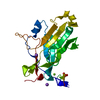
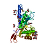
- PDB-4yii: Structure of an APC2-UBCH10 complex reveals distinctive cullin-RI... -
+ Open data

- Basic information
| Entry | Database: PDB / ID: 4yii | ||||||
|---|---|---|---|---|---|---|---|
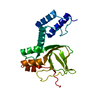
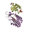
| Title | Structure of an APC2-UBCH10 complex reveals distinctive cullin-RING ligase mechanism for Anaphase-promoting complex/Cyclosome | ||||||
 Components Components |
| ||||||
 Keywords Keywords | ligase/cell cycle / ligase-cell cycle complex | ||||||
| Function / homology |  Function and homology information Function and homology informationpositive regulation of exit from mitosis / free ubiquitin chain polymerization / positive regulation of synapse maturation / Conversion from APC/C:Cdc20 to APC/C:Cdh1 in late anaphase / Inactivation of APC/C via direct inhibition of the APC/C complex / APC/C:Cdc20 mediated degradation of mitotic proteins /  anaphase-promoting complex / Aberrant regulation of mitotic exit in cancer due to RB1 defects / regulation of meiotic cell cycle / metaphase/anaphase transition of mitotic cell cycle ...positive regulation of exit from mitosis / free ubiquitin chain polymerization / positive regulation of synapse maturation / Conversion from APC/C:Cdc20 to APC/C:Cdh1 in late anaphase / Inactivation of APC/C via direct inhibition of the APC/C complex / APC/C:Cdc20 mediated degradation of mitotic proteins / anaphase-promoting complex / Aberrant regulation of mitotic exit in cancer due to RB1 defects / regulation of meiotic cell cycle / metaphase/anaphase transition of mitotic cell cycle / anaphase-promoting complex-dependent catabolic process / (E3-independent) E2 ubiquitin-conjugating enzyme / positive regulation of synaptic plasticity / Phosphorylation of the APC/C / positive regulation of mitotic metaphase/anaphase transition / positive regulation of ubiquitin protein ligase activity / exit from mitosis / protein K11-linked ubiquitination / regulation of mitotic metaphase/anaphase transition / positive regulation of dendrite morphogenesis / E2 ubiquitin-conjugating enzyme / ubiquitin-like protein ligase binding / Regulation of APC/C activators between G1/S and early anaphase / ubiquitin conjugating enzyme activity / Transcriptional Regulation by VENTX / positive regulation of axon extension / protein K48-linked ubiquitination / ubiquitin ligase complex / APC/C:Cdc20 mediated degradation of Cyclin B / regulation of mitotic cell cycle / APC-Cdc20 mediated degradation of Nek2A / Synthesis of active ubiquitin: roles of E1 and E2 enzymes / Autodegradation of Cdh1 by Cdh1:APC/C / APC/C:Cdc20 mediated degradation of Securin / Assembly of the pre-replicative complex / Cdc20:Phospho-APC/C mediated degradation of Cyclin A / APC/C:Cdh1 mediated degradation of Cdc20 and other APC/C:Cdh1 targeted proteins in late mitosis/early G1 / CDK-mediated phosphorylation and removal of Cdc6 / protein polyubiquitination / Separation of Sister Chromatids / ubiquitin-protein transferase activity / ubiquitin protein ligase activity / Antigen processing: Ubiquitination & Proteasome degradation / nervous system development / Senescence-Associated Secretory Phenotype (SASP) / ubiquitin-dependent protein catabolic process / cell differentiation / protein ubiquitination / cell division / negative regulation of gene expression / ubiquitin protein ligase binding / nucleoplasm / ATP binding / nucleus / plasma membrane / cytosol anaphase-promoting complex / Aberrant regulation of mitotic exit in cancer due to RB1 defects / regulation of meiotic cell cycle / metaphase/anaphase transition of mitotic cell cycle ...positive regulation of exit from mitosis / free ubiquitin chain polymerization / positive regulation of synapse maturation / Conversion from APC/C:Cdc20 to APC/C:Cdh1 in late anaphase / Inactivation of APC/C via direct inhibition of the APC/C complex / APC/C:Cdc20 mediated degradation of mitotic proteins / anaphase-promoting complex / Aberrant regulation of mitotic exit in cancer due to RB1 defects / regulation of meiotic cell cycle / metaphase/anaphase transition of mitotic cell cycle / anaphase-promoting complex-dependent catabolic process / (E3-independent) E2 ubiquitin-conjugating enzyme / positive regulation of synaptic plasticity / Phosphorylation of the APC/C / positive regulation of mitotic metaphase/anaphase transition / positive regulation of ubiquitin protein ligase activity / exit from mitosis / protein K11-linked ubiquitination / regulation of mitotic metaphase/anaphase transition / positive regulation of dendrite morphogenesis / E2 ubiquitin-conjugating enzyme / ubiquitin-like protein ligase binding / Regulation of APC/C activators between G1/S and early anaphase / ubiquitin conjugating enzyme activity / Transcriptional Regulation by VENTX / positive regulation of axon extension / protein K48-linked ubiquitination / ubiquitin ligase complex / APC/C:Cdc20 mediated degradation of Cyclin B / regulation of mitotic cell cycle / APC-Cdc20 mediated degradation of Nek2A / Synthesis of active ubiquitin: roles of E1 and E2 enzymes / Autodegradation of Cdh1 by Cdh1:APC/C / APC/C:Cdc20 mediated degradation of Securin / Assembly of the pre-replicative complex / Cdc20:Phospho-APC/C mediated degradation of Cyclin A / APC/C:Cdh1 mediated degradation of Cdc20 and other APC/C:Cdh1 targeted proteins in late mitosis/early G1 / CDK-mediated phosphorylation and removal of Cdc6 / protein polyubiquitination / Separation of Sister Chromatids / ubiquitin-protein transferase activity / ubiquitin protein ligase activity / Antigen processing: Ubiquitination & Proteasome degradation / nervous system development / Senescence-Associated Secretory Phenotype (SASP) / ubiquitin-dependent protein catabolic process / cell differentiation / protein ubiquitination / cell division / negative regulation of gene expression / ubiquitin protein ligase binding / nucleoplasm / ATP binding / nucleus / plasma membrane / cytosolSimilarity search - Function | ||||||
| Biological species |  Homo sapiens (human) Homo sapiens (human) | ||||||
| Method | X-RAY DIFFRACTION / SYNCHROTRON / MOLECULAR REPLACEMENT / Resolution: 1.804 Å | ||||||
 Authors Authors | Brown, N.G. / Cho, S.E. / Schulman, B.A. | ||||||
 Citation Citation | Journal: Proc.Natl.Acad.Sci.USA / Year: 2015 Title: Crystal Structure of E2 Complex Authors: Brown, N.G. / Cho, S.E. / Schulman, B.A. | ||||||
| History |
|
- Structure visualization
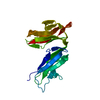
| Structure viewer | Molecule: MolmilJmol/JSmol |
|---|
- Downloads & links
-Download
| PDBx/mmCIF format | 4yii.cif.gz | 59.4 KB | Display | PDBx/mmCIF format |
|---|---|---|---|---|
| PDB format | pdb4yii.ent.gz | 40.5 KB | Display | PDB format |
| PDBx/mmJSON format | 4yii.json.gz | Tree view | PDBx/mmJSON format | |
| Others |  Other downloads Other downloads |
-Validation report
| Arichive directory | https://data.pdbj.org/pub/pdb/validation_reports/yi/4yiiftp://data.pdbj.org/pub/pdb/validation_reports/yi/4yii | HTTPS FTP |
|---|
-Related structure data


-Links
 PDBj
PDBj


- Assembly
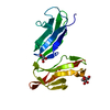
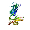
| Deposited unit | 
| ||||||||
|---|---|---|---|---|---|---|---|---|---|
| 1 |
| ||||||||
| Unit cell |
|
-Components
| #1: Protein | Mass: 17360.707 Da / Num. of mol.: 1 / Fragment: unp residues 27-179 Source method: isolated from a genetically manipulated source Source: (gene. exp.) Homo sapiens (human) / Gene: UBE2C, UBCH10 / Production host:  Escherichia coli (E. coli) / References: UniProt: O00762, ubiquitin-protein ligase Escherichia coli (E. coli) / References: UniProt: O00762, ubiquitin-protein ligase |
|---|---|
| #2: Protein | / APC2 / Cyclosome subunit 2 Mass: 10259.662 Da / Num. of mol.: 1 / Fragment: unp residues 735-822 Source method: isolated from a genetically manipulated source Source: (gene. exp.) Homo sapiens (human) / Gene: ANAPC2, APC2, KIAA1406 / Production host: Escherichia coli (E. coli) / References: UniProt: Q9UJX6 |
| #3: Water | ChemComp-HOH / Water Mass: 18.015 Da / Num. of mol.: 64 / Source method: isolated from a natural source / Formula: H2O Mass: 18.015 Da / Num. of mol.: 64 / Source method: isolated from a natural source / Formula: H2O |
-Experimental details
-Experiment
| Experiment | Method: X-RAY DIFFRACTION |
|---|
- Sample preparation
| Crystal | Density Matthews: 2.05 Å3/Da / Density % sol: 39.87 % |
|---|---|
| Crystal grow | Temperature: 296 K / Method: vapor diffusion, hanging drop / Details: PEG 3000, MES / PH range: 6.5 |
-Data collection
| Diffraction | Mean temperature: 100 K |
|---|---|
| Diffraction source | Source: SYNCHROTRON / Site: APS  / Beamline: 24-ID-C / Wavelength: 1.2826 Å / Beamline: 24-ID-C / Wavelength: 1.2826 Å |
| Detector | Type: DECTRIS PILATUS 6M-F / Detector: PIXEL / Date: Nov 20, 2014 |
| Radiation | Protocol: SINGLE WAVELENGTH / Monochromatic (M) / Laue (L): M / Scattering type: x-ray |
| Radiation wavelength | Wavelength: 1.2826 Å / Relative weight: 1 |
| Reflection | Resolution: 1.8→50 Å / Num. obs: 21055 / % possible obs: 94 % / Redundancy: 2.9 % / Rmerge(I) obs: 0.05 / Net I/σ(I): 28.4 |
| Reflection shell | Resolution: 1.8→1.86 Å / Rmerge(I) obs: 0.347 / Num. unique all: 21055 / % possible all: 68.2 |
- Processing
| Software |
| ||||||||||||||||||||||||||||||||||||||||||||||||||||||||
|---|---|---|---|---|---|---|---|---|---|---|---|---|---|---|---|---|---|---|---|---|---|---|---|---|---|---|---|---|---|---|---|---|---|---|---|---|---|---|---|---|---|---|---|---|---|---|---|---|---|---|---|---|---|---|---|---|---|
| Refinement | Method to determine structure: MOLECULAR REPLACEMENT Starting model: 1LDD, 1I7K Resolution: 1.804→44.272 Å / SU ML: 0.51 / Cross valid method: FREE R-VALUE / σ(F): 1.37 / Phase error: 25.71 / Stereochemistry target values: ML
| ||||||||||||||||||||||||||||||||||||||||||||||||||||||||
| Solvent computation | Shrinkage radii: 0.83 Å / VDW probe radii: 1.1 Å / Solvent model: FLAT BULK SOLVENT MODEL / Bsol: 50.613 Å2 / ksol: 0.378 e/Å3 | ||||||||||||||||||||||||||||||||||||||||||||||||||||||||
| Displacement parameters |
| ||||||||||||||||||||||||||||||||||||||||||||||||||||||||
| Refinement step | Cycle: LAST / Resolution: 1.804→44.272 Å
| ||||||||||||||||||||||||||||||||||||||||||||||||||||||||
| Refine LS restraints |
| ||||||||||||||||||||||||||||||||||||||||||||||||||||||||
| LS refinement shell |
|
