 Movie
Movie Controller

[English] 日本語
 Yorodumi
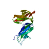
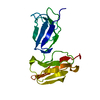
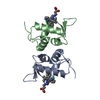
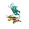
- PDB-3ry4: 1.5 Angstrom resolution structure of glycosylated fcgammariia (lo... -
+ Open data

- Basic information
| Entry | Database: PDB / ID: 3ry4 | ||||||
|---|---|---|---|---|---|---|---|
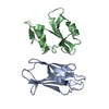
| Title | 1.5 Angstrom resolution structure of glycosylated fcgammariia (low-responder polymorphism) | ||||||
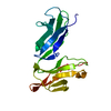
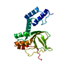
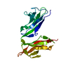
 Components Components | Low affinity immunoglobulin gamma Fc region receptor II-a | ||||||
 Keywords Keywords |  IMMUNE SYSTEM / FC RECEPTOR / CD32 / IMMUNOGLOBULIN SUPERFAMILY / LOW RESPONDER POLYMORPHISM / CELL MEMBRANE / GLYCOPROTEIN / IGG-BINDING PROTEIN / IMMUNOGLOBULIN DOMAIN / MEMBRANE / PHOSPHOPROTEIN / RECEPTOR / TRANSMEMBRANE IMMUNE SYSTEM / FC RECEPTOR / CD32 / IMMUNOGLOBULIN SUPERFAMILY / LOW RESPONDER POLYMORPHISM / CELL MEMBRANE / GLYCOPROTEIN / IGG-BINDING PROTEIN / IMMUNOGLOBULIN DOMAIN / MEMBRANE / PHOSPHOPROTEIN / RECEPTOR / TRANSMEMBRANE | ||||||
| Function / homology |  Function and homology information Function and homology informationIgG binding / immune system process / FCGR activation / regulation of immune response / Role of phospholipids in phagocytosis / FCGR3A-mediated IL10 synthesis / secretory granule membrane / Regulation of actin dynamics for phagocytic cup formation / transmembrane signaling receptor activity / cell surface receptor signaling pathway ...IgG binding / immune system process / FCGR activation / regulation of immune response / Role of phospholipids in phagocytosis / FCGR3A-mediated IL10 synthesis / secretory granule membrane / Regulation of actin dynamics for phagocytic cup formation / transmembrane signaling receptor activity / cell surface receptor signaling pathway / Neutrophil degranulation / plasma membraneSimilarity search - Function | ||||||
| Biological species |  Homo sapiens (human) Homo sapiens (human) | ||||||
| Method | X-RAY DIFFRACTION / SYNCHROTRON / MOLECULAR REPLACEMENT / Resolution: 1.5 Å | ||||||
 Authors Authors | Ramsland, P.A. / Farrugia, W. / Hogarth, P.M. | ||||||
 Citation Citation | Journal: J.Immunol. / Year: 2011 Title: Structural Basis for Fc{gamma}RIIa Recognition of Human IgG and Formation of Inflammatory Signaling Complexes. Authors: Ramsland, P.A. / Farrugia, W. / Bradford, T.M. / Sardjono, C.T. / Esparon, S. / Trist, H.M. / Powell, M.S. / Tan, P.S. / Cendron, A.C. / Wines, B.D. / Scott, A.M. / Hogarth, P.M. | ||||||
| History |
|
- Structure visualization
| Structure viewer | Molecule: MolmilJmol/JSmol |
|---|
- Downloads & links
-Download
| PDBx/mmCIF format | 3ry4.cif.gz | 55.8 KB | Display | PDBx/mmCIF format |
|---|---|---|---|---|
| PDB format | pdb3ry4.ent.gz | 38.2 KB | Display | PDB format |
| PDBx/mmJSON format | 3ry4.json.gz | Tree view | PDBx/mmJSON format | |
| Others |  Other downloads Other downloads |
-Validation report
| Arichive directory | https://data.pdbj.org/pub/pdb/validation_reports/ry/3ry4ftp://data.pdbj.org/pub/pdb/validation_reports/ry/3ry4 | HTTPS FTP |
|---|
-Related structure data
| Related structure data |  3ry5C 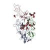 3ry6C  1fcgS C: citing same article ( S: Starting model for refinement |
|---|---|
| Similar structure data |

-Links
 PDBj
PDBj


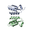
- Assembly
| Deposited unit | 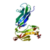
| |||||||||
|---|---|---|---|---|---|---|---|---|---|---|
| 1 |
| |||||||||
| Unit cell |
| |||||||||
| Components on special symmetry positions |
|
-Components
| #1: Protein | Mass: 19187.449 Da / Num. of mol.: 1 / Fragment: EXTRACELLULAR DOMAIN, residues 37-206 / Mutation: S88P Source method: isolated from a genetically manipulated source Source: (gene. exp.) Homo sapiens (human) / Tissue: Blood / Gene: CD32, FCG2, FCGR2A, FCGR2A1, IGFR2 / Plasmid: PVL1392 / Cell line (production host): SF21 / Production host:   Spodoptera frugiperda (fall armyworm) / References: UniProt: P12318 Spodoptera frugiperda (fall armyworm) / References: UniProt: P12318 |
|---|---|
| #2: Sugar | ChemComp-NAG / N-Acetylglucosamine  Type: D-saccharide, beta linking / Mass: 221.208 Da / Num. of mol.: 1 Type: D-saccharide, beta linking / Mass: 221.208 Da / Num. of mol.: 1Source method: isolated from a genetically manipulated source Formula: C8H15NO6 |
| #3: Chemical | ChemComp-GOL / Glycerol  Mass: 92.094 Da / Num. of mol.: 1 / Source method: obtained synthetically / Formula: C3H8O3 Mass: 92.094 Da / Num. of mol.: 1 / Source method: obtained synthetically / Formula: C3H8O3 |
| #4: Water | ChemComp-HOH / Water Mass: 18.015 Da / Num. of mol.: 267 / Source method: isolated from a natural source / Formula: H2O Mass: 18.015 Da / Num. of mol.: 267 / Source method: isolated from a natural source / Formula: H2O |
-Experimental details
-Experiment
| Experiment | Method: X-RAY DIFFRACTION / Number of used crystals: 1 |
|---|
- Sample preparation
| Crystal | Density Matthews: 2.9 Å3/Da / Density % sol: 57.55 % |
|---|---|
| Crystal grow | Method: vapor diffusion / pH: 5.6 Details: 30% (W/V) PEG 4000, 0.2M AMMONIUM ACETATE, 0.1M SODIUM CITRATE, PH 5.60, VAPOR DIFFUSION, TEMPERATURE 291.0K |
-Data collection
| Diffraction | Mean temperature: 100 K |
|---|---|
| Diffraction source | Source: SYNCHROTRON / Site: APS  / Beamline: 14-BM-C / Wavelength: 0.9 / Beamline: 14-BM-C / Wavelength: 0.9 |
| Detector | Type: ADSC QUANTUM 4 / Detector: CCD / Date: Apr 11, 2002 / Details: BENT CONICAL SI-MIRROR (RH COATED) |
| Radiation | Monochromator: BENT GE(111) MONOCHROMATOR / Protocol: SINGLE WAVELENGTH / Monochromatic (M) / Laue (L): M / Scattering type: x-ray |
| Radiation wavelength | Wavelength: 0.9 Å / Relative weight: 1 |
| Reflection | Resolution: 1.5→20 Å / Num. obs: 35275 / % possible obs: 94.5 % / Observed criterion σ(I): 0 / Redundancy: 6.1 % / Biso Wilson estimate: 20.3 Å2 / Rsym value: 0.052 / Net I/σ(I): 21.8 |
| Reflection shell | Resolution: 1.5→1.55 Å / Redundancy: 4 % / Mean I/σ(I) obs: 3.8 / Rsym value: 0.32 / % possible all: 85.5 |
- Processing
| Software |
| ||||||||||||||||||||||||||||||||||||||||||||||||||||||||||||
|---|---|---|---|---|---|---|---|---|---|---|---|---|---|---|---|---|---|---|---|---|---|---|---|---|---|---|---|---|---|---|---|---|---|---|---|---|---|---|---|---|---|---|---|---|---|---|---|---|---|---|---|---|---|---|---|---|---|---|---|---|---|
| Refinement | Method to determine structure: MOLECULAR REPLACEMENT Starting model: 1FCG Resolution: 1.5→19.84 Å / Cross valid method: THROUGHOUT / σ(F): 0 / Stereochemistry target values: ENGH & HUBER
| ||||||||||||||||||||||||||||||||||||||||||||||||||||||||||||
| Refine analyze |
| ||||||||||||||||||||||||||||||||||||||||||||||||||||||||||||
| Refinement step | Cycle: LAST / Resolution: 1.5→19.84 Å
| ||||||||||||||||||||||||||||||||||||||||||||||||||||||||||||
| Refine LS restraints |
| ||||||||||||||||||||||||||||||||||||||||||||||||||||||||||||
| LS refinement shell | Resolution: 1.5→1.55 Å / Rfactor Rfree error: 0.031 /
|
