 Movie
Movie Controller


+ Open data

- Basic information
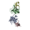
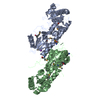
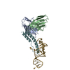
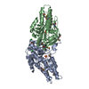
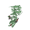
| Entry | Database: PDB / ID: 1fcg | ||||||
|---|---|---|---|---|---|---|---|
| Title | ECTODOMAIN OF HUMAN FC GAMMA RECEPTOR, FCGRIIA | ||||||
 Components Components | PROTEIN (FC RECEPTOR FC(GAMMA)RIIA) | ||||||
 Keywords Keywords |  IMMUNE SYSTEM / MEMBRANE PROTEIN / FC RECEPTOR / IMMUNOGLOULIN / LEUKOCYTE / CD32 IMMUNE SYSTEM / MEMBRANE PROTEIN / FC RECEPTOR / IMMUNOGLOULIN / LEUKOCYTE / CD32 | ||||||
| Function / homology |  Function and homology information Function and homology informationIgG binding / immune system process / FCGR activation / regulation of immune response / Role of phospholipids in phagocytosis / FCGR3A-mediated IL10 synthesis / secretory granule membrane / Regulation of actin dynamics for phagocytic cup formation / transmembrane signaling receptor activity / cell surface receptor signaling pathway ...IgG binding / immune system process / FCGR activation / regulation of immune response / Role of phospholipids in phagocytosis / FCGR3A-mediated IL10 synthesis / secretory granule membrane / Regulation of actin dynamics for phagocytic cup formation / transmembrane signaling receptor activity / cell surface receptor signaling pathway / Neutrophil degranulation / plasma membraneSimilarity search - Function | ||||||
| Biological species |  Homo sapiens (human) Homo sapiens (human) | ||||||
| Method | X-RAY DIFFRACTION / SIRAS / Resolution: 2 Å | ||||||
 Authors Authors | Maxwell, K.F. / Powell, M.S. / Garrett, T.P. / Hogarth, P.M. | ||||||
 Citation Citation | Journal: Nat.Struct.Biol. / Year: 1999 Title: Crystal structure of the human leukocyte Fc receptor, Fc gammaRIIa. Authors: Maxwell, K.F. / Powell, M.S. / Hulett, M.D. / Barton, P.A. / McKenzie, I.F. / Garrett, T.P. / Hogarth, P.M. #1: Journal: Immunol.Lett. / Year: 1999Title: Biochemical Analysis and Crystallisation of Fc(Gamma)RIIA, the Low Affinity Receptor for IgG Authors: Powell, M.S. / Barton, P.A. / Emmanouilidis, D. / Wines, B.D. / Neumann, G.M. / Peitersz, G.A. / Maxwell, K.F. / Garrett, T.P. / Hogarth, P.M. | ||||||
| History |
|
- Structure visualization
| Structure viewer | Molecule: MolmilJmol/JSmol |
|---|
- Downloads & links
-Download
| PDBx/mmCIF format | 1fcg.cif.gz | 48.9 KB | Display | PDBx/mmCIF format |
|---|---|---|---|---|
| PDB format | pdb1fcg.ent.gz | 35.4 KB | Display | PDB format |
| PDBx/mmJSON format | 1fcg.json.gz | Tree view | PDBx/mmJSON format | |
| Others |  Other downloads Other downloads |
-Validation report
| Arichive directory | https://data.pdbj.org/pub/pdb/validation_reports/fc/1fcgftp://data.pdbj.org/pub/pdb/validation_reports/fc/1fcg | HTTPS FTP |
|---|
-Related structure data
| Similar structure data |
|---|


-Links
 PDBj
PDBj


- Assembly
| Deposited unit | 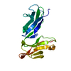
| ||||||||
|---|---|---|---|---|---|---|---|---|---|
| 1 | 
| ||||||||
| Unit cell |
| ||||||||
| Components on special symmetry positions |
|
-Components
| #1: Protein | Mass: 19556.863 Da / Num. of mol.: 1 / Fragment: EXTRACELLULAR DOMAINS / Mutation: S88F, R134H Source method: isolated from a genetically manipulated source Details: LOW RESPONDER VARIANT OF FC(GAMMA)RIIA / Source: (gene. exp.) Homo sapiens (human) / Tissue: BLOOD / Cell: MONOCYTE / Cellular location: CELL SURFACECell membrane / Plasmid: PVL1392 / Cell line (production host): SF21 / Cellular location (production host): SECRETED / Production host:   Spodoptera frugiperda (fall armyworm) / References: UniProt: P12318 Spodoptera frugiperda (fall armyworm) / References: UniProt: P12318 |
|---|---|
| #2: Water | ChemComp-HOH / Water Mass: 18.015 Da / Num. of mol.: 91 / Source method: isolated from a natural source / Formula: H2O Mass: 18.015 Da / Num. of mol.: 91 / Source method: isolated from a natural source / Formula: H2O |
-Experimental details
-Experiment
| Experiment | Method: X-RAY DIFFRACTION / Number of used crystals: 1 |
|---|
- Sample preparation
| Crystal | Density Matthews: 2.6 Å3/Da / Density % sol: 53 % | |||||||||||||||||||||||||
|---|---|---|---|---|---|---|---|---|---|---|---|---|---|---|---|---|---|---|---|---|---|---|---|---|---|---|
| Crystal grow | pH: 5.6 Details: HANGING DROP VAPOUR DIFFUSION WITH EQUAL VOLUMES OF FC(GAMMA)RIIA AT 6 MG/ML AND MOTHER LIQUOR (0.2 M AMMONIUM ACETATE, 30% W/V PEG 4000, 0.1 M SODIUM CITRATE PH 5.6) MIXED IN 3 MICRO L ...Details: HANGING DROP VAPOUR DIFFUSION WITH EQUAL VOLUMES OF FC(GAMMA)RIIA AT 6 MG/ML AND MOTHER LIQUOR (0.2 M AMMONIUM ACETATE, 30% W/V PEG 4000, 0.1 M SODIUM CITRATE PH 5.6) MIXED IN 3 MICRO L DROPLETS AND ALLOWED TO EQUILIBRATE AT 22 C FOR 3-9 DAYS. | |||||||||||||||||||||||||
| Crystal | *PLUS | |||||||||||||||||||||||||
| Crystal grow | *PLUS Temperature: 22 ℃ / Method: vapor diffusion, hanging drop / Details: Powell, M.S., (1999) Immunol.Lett., 68, 17. | |||||||||||||||||||||||||
| Components of the solutions | *PLUS
|
-Data collection
| Diffraction | Mean temperature: 108 K |
|---|---|
| Diffraction source | Source: ROTATING ANODE / Type: MACSCIENCE M18X / Wavelength: 1.5418 |
| Detector | Type: RIGAKU / Detector: IMAGE PLATE / Date: Apr 1, 1998 / Details: MIRRORS |
| Radiation | Monochromator: NI FILTER / Protocol: SINGLE WAVELENGTH / Monochromatic (M) / Laue (L): M / Scattering type: x-ray |
| Radiation wavelength | Wavelength: 1.5418 Å / Relative weight: 1 |
| Reflection | Resolution: 2→10 Å / Num. obs: 15024 / % possible obs: 94.9 % / Redundancy: 2.73 % / Rmerge(I) obs: 0.069 / Net I/σ(I): 10.3 |
| Reflection shell | Resolution: 2→2.07 Å / Redundancy: 2.53 % / Rmerge(I) obs: 0.359 / Mean I/σ(I) obs: 1.5 / % possible all: 95.9 |
| Reflection shell | *PLUS Highest resolution: 2 Å / % possible obs: 95.9 % |
- Processing
| Software |
| ||||||||||||||||||||||||||||||||||||||||||||||||||||||||||||||||||||||||||||||||||||
|---|---|---|---|---|---|---|---|---|---|---|---|---|---|---|---|---|---|---|---|---|---|---|---|---|---|---|---|---|---|---|---|---|---|---|---|---|---|---|---|---|---|---|---|---|---|---|---|---|---|---|---|---|---|---|---|---|---|---|---|---|---|---|---|---|---|---|---|---|---|---|---|---|---|---|---|---|---|---|---|---|---|---|---|---|---|
| Refinement | Method to determine structure: SIRAS / Resolution: 2→6 Å / SU B: 4.65 / SU ML: 0.13 / Cross valid method: THROUGHOUT / σ(F): 0 / ESU R: 0.21 / ESU R Free: 0.18 Details: NO ELECTRON DENSITY WAS OBSERVED FOR AMINO ACIDS 1-3 OR EITHER OF THE N-LINKED CARBOHYDRATE MOIETIES SO THESE RESIDUES HAVE NOT BEEN MODELLED. MASS SPECTROMETRY INDICATES THAT THESE RESIDUES ...Details: NO ELECTRON DENSITY WAS OBSERVED FOR AMINO ACIDS 1-3 OR EITHER OF THE N-LINKED CARBOHYDRATE MOIETIES SO THESE RESIDUES HAVE NOT BEEN MODELLED. MASS SPECTROMETRY INDICATES THAT THESE RESIDUES ARE PRESENT IN THE CRYSTAL.
| ||||||||||||||||||||||||||||||||||||||||||||||||||||||||||||||||||||||||||||||||||||
| Displacement parameters | Biso mean: 25.1 Å2 | ||||||||||||||||||||||||||||||||||||||||||||||||||||||||||||||||||||||||||||||||||||
| Refinement step | Cycle: LAST / Resolution: 2→6 Å
| ||||||||||||||||||||||||||||||||||||||||||||||||||||||||||||||||||||||||||||||||||||
| Refine LS restraints |
| ||||||||||||||||||||||||||||||||||||||||||||||||||||||||||||||||||||||||||||||||||||
| Software | *PLUS Name: REFMAC / Classification: refinement | ||||||||||||||||||||||||||||||||||||||||||||||||||||||||||||||||||||||||||||||||||||
| Refinement | *PLUS Highest resolution: 2 Å / σ(F): 0 / % reflection Rfree: 8 % | ||||||||||||||||||||||||||||||||||||||||||||||||||||||||||||||||||||||||||||||||||||
| Solvent computation | *PLUS | ||||||||||||||||||||||||||||||||||||||||||||||||||||||||||||||||||||||||||||||||||||
| Displacement parameters | *PLUS Biso mean: 25.1 Å2 | ||||||||||||||||||||||||||||||||||||||||||||||||||||||||||||||||||||||||||||||||||||
| Refine LS restraints | *PLUS
|
