 Movie
Movie Controller


+ Open data

- Basic information
| Entry | Database: PDB / ID: 1e4j | ||||||
|---|---|---|---|---|---|---|---|
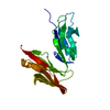
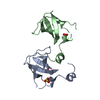
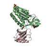
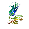
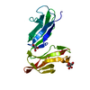
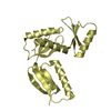
| Title | Crystal structure of the soluble human Fc-gamma Receptor III | ||||||
 Components Components | LOW AFFINITY IMMUNOGLOBULIN GAMMA FC RECEPTOR III | ||||||
 Keywords Keywords |  IMMUNE SYSTEM / IGG / FC / RECEPTOR / CD16 / GAMMA IMMUNE SYSTEM / IGG / FC / RECEPTOR / CD16 / GAMMA | ||||||
| Function / homology |  Function and homology informationGPI anchor binding / Post-translational modification: synthesis of GPI-anchored proteins / IgG binding / regulation of immune response / side of membrane / secretory granule membrane / transmembrane signaling receptor activity / cell surface receptor signaling pathway / immune response / Neutrophil degranulation ...GPI anchor binding / Post-translational modification: synthesis of GPI-anchored proteins / IgG binding / regulation of immune response / side of membrane / secretory granule membrane / transmembrane signaling receptor activity / cell surface receptor signaling pathway / immune response / Neutrophil degranulation / extracellular exosome / extracellular region / plasma membrane Function and homology informationGPI anchor binding / Post-translational modification: synthesis of GPI-anchored proteins / IgG binding / regulation of immune response / side of membrane / secretory granule membrane / transmembrane signaling receptor activity / cell surface receptor signaling pathway / immune response / Neutrophil degranulation ...GPI anchor binding / Post-translational modification: synthesis of GPI-anchored proteins / IgG binding / regulation of immune response / side of membrane / secretory granule membrane / transmembrane signaling receptor activity / cell surface receptor signaling pathway / immune response / Neutrophil degranulation / extracellular exosome / extracellular region / plasma membraneSimilarity search - Function | ||||||
| Biological species |  HOMO SAPIENS (human) HOMO SAPIENS (human) | ||||||
| Method | X-RAY DIFFRACTION / MOLECULAR REPLACEMENT / Resolution: 2.5 Å | ||||||
 Authors Authors | Sondermann, P. / Huber, R. / Jacob, U. | ||||||
 Citation Citation | Journal: Nature / Year: 2000 Title: The 3.2-A Crystal Structure of the Human Igg1 Fc Fragment-Fc Gammariii Complex. Authors: Sondermann, P. / Huber, R. / Oosthuizen, V. / Jacob, U. | ||||||
| History |
|
- Structure visualization
| Structure viewer | Molecule: MolmilJmol/JSmol |
|---|
- Downloads & links
-Download
| PDBx/mmCIF format | 1e4j.cif.gz | 48.8 KB | Display | PDBx/mmCIF format |
|---|---|---|---|---|
| PDB format | pdb1e4j.ent.gz | 33.8 KB | Display | PDB format |
| PDBx/mmJSON format | 1e4j.json.gz | Tree view | PDBx/mmJSON format | |
| Others |  Other downloads Other downloads |
-Validation report
| Arichive directory | https://data.pdbj.org/pub/pdb/validation_reports/e4/1e4jftp://data.pdbj.org/pub/pdb/validation_reports/e4/1e4j | HTTPS FTP |
|---|
-Related structure data
| Related structure data |  1e4kC 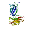 2fcbS S: Starting model for refinement C: citing same article ( |
|---|---|
| Similar structure data |


-Links
 PDBj
PDBj
- Assembly
| Deposited unit | 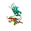
| ||||||||
|---|---|---|---|---|---|---|---|---|---|
| 1 |
| ||||||||
| Unit cell |
|
-Components
| #1: Protein | Mass: 20121.453 Da / Num. of mol.: 1 / Fragment: EXTRACELLULAR DOMAIN Source method: isolated from a genetically manipulated source Source: (gene. exp.) HOMO SAPIENS (human) / Cell: LEUKOCYTE / Plasmid: PET21 / Cellular location (production host): INCLUSION BODIES / Production host:  ESCHERICHIA COLI (E. coli) / Strain (production host): BL21(DE3) / References: UniProt: O75015 ESCHERICHIA COLI (E. coli) / Strain (production host): BL21(DE3) / References: UniProt: O75015 |
|---|---|
| #2: Water | ChemComp-HOH / Water Mass: 18.015 Da / Num. of mol.: 67 / Source method: isolated from a natural source / Formula: H2O Mass: 18.015 Da / Num. of mol.: 67 / Source method: isolated from a natural source / Formula: H2O |
-Experimental details
-Experiment
| Experiment | Method: X-RAY DIFFRACTION / Number of used crystals: 1 |
|---|
- Sample preparation
| Crystal | Density Matthews: 2.37 Å3/Da / Density % sol: 48 % | ||||||||||||||||||||||||||||||||||||||||||||||||||||||
|---|---|---|---|---|---|---|---|---|---|---|---|---|---|---|---|---|---|---|---|---|---|---|---|---|---|---|---|---|---|---|---|---|---|---|---|---|---|---|---|---|---|---|---|---|---|---|---|---|---|---|---|---|---|---|---|
| Crystal grow | pH: 8 / Details: 0.1M MES/TRIS PH 7.8, 22.0% PEG8000 | ||||||||||||||||||||||||||||||||||||||||||||||||||||||
| Crystal grow | *PLUS Temperature: 18 ℃ / pH: 7.1 / Method: vapor diffusion, sitting drop | ||||||||||||||||||||||||||||||||||||||||||||||||||||||
| Components of the solutions | *PLUS
|
-Data collection
| Diffraction | Mean temperature: 291 K |
|---|---|
| Diffraction source | Source: ROTATING ANODE / Type: RIGAKU RU200 / Wavelength: 1.5418 |
| Detector | Type: MARRESEARCH / Detector: IMAGE PLATE / Date: Sep 15, 1999 |
| Radiation | Monochromator: NI FILTER / Protocol: SINGLE WAVELENGTH / Monochromatic (M) / Laue (L): M / Scattering type: x-ray |
| Radiation wavelength | Wavelength: 1.5418 Å / Relative weight: 1 |
| Reflection | Resolution: 2.5→40 Å / Num. obs: 6725 / % possible obs: 94.7 % / Observed criterion σ(I): 2 / Redundancy: 3 % / Rmerge(I) obs: 0.114 |
| Reflection shell | Resolution: 2.3→2.5 Å / % possible all: 50 |
| Reflection shell | *PLUS % possible obs: 72.3 % / Rmerge(I) obs: 0.5 |
- Processing
| Software |
| ||||||||||||||||||||||||||||||||||||||||||||||||||||||||||||
|---|---|---|---|---|---|---|---|---|---|---|---|---|---|---|---|---|---|---|---|---|---|---|---|---|---|---|---|---|---|---|---|---|---|---|---|---|---|---|---|---|---|---|---|---|---|---|---|---|---|---|---|---|---|---|---|---|---|---|---|---|---|
| Refinement | Method to determine structure: MOLECULAR REPLACEMENT Starting model: 2FCB Resolution: 2.5→50 Å / Cross valid method: THROUGHOUT
| ||||||||||||||||||||||||||||||||||||||||||||||||||||||||||||
| Solvent computation | Solvent model: DENSITY MODIFICATION | ||||||||||||||||||||||||||||||||||||||||||||||||||||||||||||
| Displacement parameters | Biso mean: 29.7 Å2
| ||||||||||||||||||||||||||||||||||||||||||||||||||||||||||||
| Refinement step | Cycle: LAST / Resolution: 2.5→50 Å
| ||||||||||||||||||||||||||||||||||||||||||||||||||||||||||||
| Refine LS restraints |
| ||||||||||||||||||||||||||||||||||||||||||||||||||||||||||||
| Xplor file |
|
