[English] 日本語
Yorodumi
- PDB-4xi9: Human OGT in complex with UDP-5S-GlcNAc and substrate peptide (RBL2) -

+
Open data


ID or keywords:

Loading...

-
Basic information

Entry
Database: PDB / ID: 4xi9
TitleHuman OGT in complex with UDP-5S-GlcNAc and substrate peptide (RBL2)
Components
  • Retinoblastoma-like protein 2
  • UDP-N-acetylglucosamine--peptide N-acetylglucosaminyltransferase 110 kDa subunit
Keywords
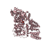
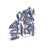
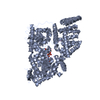
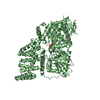
TRANSFERASE / O-GlcNAc transferase inverting GT-B substrate complex
Function / homology
Function and homology information


regulation of lipid kinase activity / protein N-acetylglucosaminyltransferase complex / protein O-GlcNAc transferase / regulation of insulin receptor signaling pathway / protein O-acetylglucosaminyltransferase activity / positive regulation of transcription from RNA polymerase II promoter by glucose / FOXO-mediated transcription of cell cycle genes / acetylglucosaminyltransferase activity / regulation of necroptotic process / regulation of Rac protein signal transduction ...regulation of lipid kinase activity / protein N-acetylglucosaminyltransferase complex / protein O-GlcNAc transferase / regulation of insulin receptor signaling pathway / protein O-acetylglucosaminyltransferase activity / positive regulation of transcription from RNA polymerase II promoter by glucose / FOXO-mediated transcription of cell cycle genes / acetylglucosaminyltransferase activity / regulation of necroptotic process / regulation of Rac protein signal transduction / negative regulation of stem cell population maintenance / Transcription of E2F targets under negative control by DREAM complex / protein O-linked glycosylation / Transcription of E2F targets under negative control by p107 (RBL1) and p130 (RBL2) in complex with HDAC1 / NSL complex / : / regulation of neurotransmitter receptor localization to postsynaptic specialization membrane / regulation of glycolytic process / RIPK1-mediated regulated necrosis / regulation of synapse assembly / regulation of gluconeogenesis / negative regulation of G1/S transition of mitotic cell cycle / G1/S-Specific Transcription / positive regulation of stem cell population maintenance / Formation of WDR5-containing histone-modifying complexes / positive regulation of proteolysis / phosphatidylinositol-3,4,5-trisphosphate binding / mitophagy / hemopoiesis / G0 and Early G1 / negative regulation of proteasomal ubiquitin-dependent protein catabolic process / histone acetyltransferase complex / positive regulation of lipid biosynthetic process / Cyclin E associated events during G1/S transition / Cyclin A:Cdk2-associated events at S phase entry / negative regulation of protein ubiquitination / positive regulation of TORC1 signaling / response to nutrient / TP53 Regulates Transcription of Genes Involved in G2 Cell Cycle Arrest / negative regulation of cell migration / positive regulation of translation / cell projection / promoter-specific chromatin binding / cellular response to glucose stimulus / mitochondrial membrane / RNA polymerase II transcription regulatory region sequence-specific DNA binding / negative regulation of transforming growth factor beta receptor signaling pathway / circadian regulation of gene expression / response to insulin / Regulation of necroptotic cell death / chromatin DNA binding / protein processing / UCH proteinases / Cyclin D associated events in G1 / chromosome / chromatin organization / positive regulation of cold-induced thermogenesis / HATs acetylate histones / transcription regulator complex / cell differentiation / cell cycle / negative regulation of gene expression / glutamatergic synapse / apoptotic process / chromatin / nucleolus / regulation of transcription by RNA polymerase II / positive regulation of DNA-templated transcription / negative regulation of transcription by RNA polymerase II / signal transduction / positive regulation of transcription by RNA polymerase II / protein-containing complex / extracellular exosome / nucleoplasm / nucleus / plasma membrane / cytosol
Similarity search - Function
Signal recognition particle alu RNA binding heterodimer, srp9/1 - #150 / Retinoblastoma-associated protein, B-box / Retinoblastoma-associated protein, A-box / Retinoblastoma-associated protein, C-terminal / Retinoblastoma-associated protein, N-terminal / Retinoblastoma protein family / Retinoblastoma-associated protein B domain / Retinoblastoma-associated protein A domain / Domain of unknown function (DUF3452) / Domain of unknown function (DUF3452) ...Signal recognition particle alu RNA binding heterodimer, srp9/1 - #150 / Retinoblastoma-associated protein, B-box / Retinoblastoma-associated protein, A-box / Retinoblastoma-associated protein, C-terminal / Retinoblastoma-associated protein, N-terminal / Retinoblastoma protein family / Retinoblastoma-associated protein B domain / Retinoblastoma-associated protein A domain / Domain of unknown function (DUF3452) / Domain of unknown function (DUF3452) / Retinoblastoma-associated protein A domain / Rb C-terminal domain / Rossmann fold - #11380 / Signal recognition particle alu RNA binding heterodimer, srp9/1 / O-GlcNAc transferase, C-terminal / Glycosyl transferase family 41 / TPR repeat / Tetratricopeptide repeat / Tetratricopeptide repeat 1 / Tetratricopeptide repeat / Tetratricopeptide repeat domain / Tetratricopeptide repeat / Glycogen Phosphorylase B; / Cyclin-like / domain present in cyclins, TFIIB and Retinoblastoma / Cyclin-like superfamily / TPR repeat region circular profile. / TPR repeat profile. / Tetratricopeptide repeats / Tetratricopeptide repeat / Serine Threonine Protein Phosphatase 5, Tetratricopeptide repeat / Alpha Horseshoe / Tetratricopeptide-like helical domain superfamily / Rossmann fold / 2-Layer Sandwich / 3-Layer(aba) Sandwich / Mainly Alpha / Alpha Beta
Similarity search - Domain/homology
Chem-12V / UDP-N-acetylglucosamine--peptide N-acetylglucosaminyltransferase 110 kDa subunit / Retinoblastoma-like protein 2
Similarity search - Component
Biological speciesHomo sapiens (human)
MethodX-RAY DIFFRACTION / SYNCHROTRON / Resolution: 3.1 Å
AuthorsSchimpl, M. / van Aalten, D.M.F.
Funding support United Kingdom, 1items
OrganizationGrant numberCountry
Wellcome TrustWT087590MA United Kingdom
CitationJournal: Nat.Struct.Mol.Biol. / Year: 2015
Title: The active site of O-GlcNAc transferase imposes constraints on substrate sequence.
Authors: Pathak, S. / Alonso, J. / Schimpl, M. / Rafie, K. / Blair, D.E. / Borodkin, V.S. / Schuttelkopf, A.W. / Albarbarawi, O. / van Aalten, D.M.
History
DepositionJan 6, 2015Deposition site: RCSB / Processing site: PDBE
Revision 1.0Aug 5, 2015Provider: repository / Type: Initial release
Revision 1.1Aug 12, 2015Group: Database references
Revision 1.2Sep 16, 2015Group: Database references

-
Structure visualization

Structure viewerMolecule:
MolmilJmol/JSmol

Downloads & links

-
Assembly

Deposited unit
A: UDP-N-acetylglucosamine--peptide N-acetylglucosaminyltransferase 110 kDa subunit
B: UDP-N-acetylglucosamine--peptide N-acetylglucosaminyltransferase 110 kDa subunit
C: UDP-N-acetylglucosamine--peptide N-acetylglucosaminyltransferase 110 kDa subunit
D: UDP-N-acetylglucosamine--peptide N-acetylglucosaminyltransferase 110 kDa subunit
E: Retinoblastoma-like protein 2
F: Retinoblastoma-like protein 2
G: Retinoblastoma-like protein 2
H: Retinoblastoma-like protein 2
hetero molecules


Theoretical massNumber of molelcules
Total (without water)329,37112
Polymers326,8778
Non-polymers2,4944
Water0
1
A: UDP-N-acetylglucosamine--peptide N-acetylglucosaminyltransferase 110 kDa subunit
E: Retinoblastoma-like protein 2
hetero molecules


Theoretical massNumber of molelcules
Total (without water)82,3433
Polymers81,7192
Non-polymers6231
Water0
TypeNameSymmetry operationNumber
identity operation1_555x,y,z1
2
B: UDP-N-acetylglucosamine--peptide N-acetylglucosaminyltransferase 110 kDa subunit
F: Retinoblastoma-like protein 2
hetero molecules


Theoretical massNumber of molelcules
Total (without water)82,3433
Polymers81,7192
Non-polymers6231
Water0
TypeNameSymmetry operationNumber
identity operation1_555x,y,z1
3
C: UDP-N-acetylglucosamine--peptide N-acetylglucosaminyltransferase 110 kDa subunit
G: Retinoblastoma-like protein 2
hetero molecules


Theoretical massNumber of molelcules
Total (without water)82,3433
Polymers81,7192
Non-polymers6231
Water0
TypeNameSymmetry operationNumber
identity operation1_555x,y,z1
4
D: UDP-N-acetylglucosamine--peptide N-acetylglucosaminyltransferase 110 kDa subunit
H: Retinoblastoma-like protein 2
hetero molecules


Theoretical massNumber of molelcules
Total (without water)82,3433
Polymers81,7192
Non-polymers6231
Water0
TypeNameSymmetry operationNumber
identity operation1_555x,y,z1
Unit cell
Length a, b, c (Å)273.910, 273.910, 142.930
Angle α, β, γ (deg.)90.00, 90.00, 120.00
Int Tables number150
Space group name H-MP321
Noncrystallographic symmetry (NCS)NCS domain:
IDEns-IDDetails
11A
21B
31C
41D

NCS domain segments:
Dom-IDComponent-IDEns-IDRefine codeAuth asym-IDAuth seq-ID
1111A312 - 1028
2111B312 - 1028
3111C312 - 1028
4111D312 - 1028

-
Components

#1: Protein
UDP-N-acetylglucosamine--peptide N-acetylglucosaminyltransferase 110 kDa subunit / O-GlcNAc transferase subunit p110 / O-linked N-acetylglucosamine transferase 110 kDa subunit / OGT


Mass: 80974.508 Da / Num. of mol.: 4 / Fragment: UNP residues 323-1041
Source method: isolated from a genetically manipulated source
Source: (gene. exp.) Homo sapiens (human) / Gene: OGT / Production host: Escherichia coli (E. coli) / References: UniProt: O15294, protein O-GlcNAc transferase
#2: Protein/peptide
Retinoblastoma-like protein 2 / / 130 kDa retinoblastoma-associated protein / p130 / Retinoblastoma-related protein 2 / RBR-2 / pRb2


Mass: 744.833 Da / Num. of mol.: 4 / Fragment: UNP residues 416-422 / Source method: obtained synthetically / Source: (synth.) Homo sapiens (human) / References: UniProt: Q08999
#3: Chemical
ChemComp-12V / (2S,3R,4R,5S,6R)-3-(acetylamino)-4,5-dihydroxy-6-(hydroxymethyl)tetrahydro-2H-thiopyran-2-yl [(2R,3S,4R,5R)-5-(2,4-dioxo-3,4-dihydropyrimidin-1(2H)-yl)-3,4-dihydroxytetrahydrofuran-2-yl]methyl dihydrogen diphosphate / URIDINE DIPHOSPHO-5-THIO-N-ACETYLGLUCOSAMINE


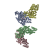
Mass: 623.419 Da / Num. of mol.: 4 / Source method: obtained synthetically / Formula: C17H27N3O16P2S

-
Experimental details

-
Experiment

ExperimentMethod: X-RAY DIFFRACTION

-
Sample preparation

CrystalDensity Matthews: 4.74 Å3/Da / Density % sol: 74.02 %
Crystal growTemperature: 293 K / Method: vapor diffusion, hanging drop
Details: 1.45 M potassium phosphate, 10 mM EDTA, 1% (w/v) xylitol

-
Data collection

DiffractionMean temperature: 100 K
Diffraction sourceSource: SYNCHROTRON / Site: Diamond / Beamline: I04-1 / Wavelength: 0.922 Å
DetectorType: PSI PILATUS 6M / Detector: PIXEL / Date: Jul 12, 2012
RadiationProtocol: SINGLE WAVELENGTH / Monochromatic (M) / Laue (L): M / Scattering type: x-ray
Radiation wavelengthWavelength: 0.922 Å / Relative weight: 1
ReflectionResolution: 3.1→25 Å / Num. obs: 108153 / % possible obs: 99.7 % / Redundancy: 3.8 % / Net I/σ(I): 9.1

-
Processing

Software
NameVersionClassification
REFMAC5.5.0088refinement
SCALEPACKdata scaling
MOSFLMdata reduction
MOLREPphasing
RefinementResolution: 3.1→25 Å / Cor.coef. Fo:Fc: 0.906 / Cor.coef. Fo:Fc free: 0.892 / SU B: 14.547 / SU ML: 0.247 / Cross valid method: THROUGHOUT / ESU R: 0.697 / ESU R Free: 0.318 / Stereochemistry target values: MAXIMUM LIKELIHOOD / Details: HYDROGENS HAVE BEEN ADDED IN THE RIDING POSITIONS
RfactorNum. reflection% reflectionSelection details
Rfree0.22346 2196 2 %RANDOM
Rwork0.20264 ---
obs0.20307 108153 99.07 %-
Solvent computationIon probe radii: 0.8 Å / Shrinkage radii: 0.8 Å / VDW probe radii: 1.4 Å / Solvent model: MASK
Displacement parametersBiso mean: 36.873 Å2
Baniso -1Baniso -2Baniso -3
1-1.02 Å20.51 Å2-0 Å2
2--1.02 Å2-0 Å2
3----1.53 Å2
Refinement stepCycle: LAST / Resolution: 3.1→25 Å
ProteinNucleic acidLigandSolventTotal
Num. atoms22244 0 156 0 22400
Refine LS restraints
Refine-IDTypeDev idealDev ideal targetNumber
X-RAY DIFFRACTIONr_bond_refined_d0.0130.02222916
X-RAY DIFFRACTIONr_bond_other_d
X-RAY DIFFRACTIONr_angle_refined_deg1.3521.96531112
X-RAY DIFFRACTIONr_angle_other_deg
X-RAY DIFFRACTIONr_dihedral_angle_1_deg5.64652804
X-RAY DIFFRACTIONr_dihedral_angle_2_deg39.16624.5591044
X-RAY DIFFRACTIONr_dihedral_angle_3_deg17.33153924
X-RAY DIFFRACTIONr_dihedral_angle_4_deg17.82115120
X-RAY DIFFRACTIONr_chiral_restr0.0880.23468
X-RAY DIFFRACTIONr_gen_planes_refined0.0060.02117324
X-RAY DIFFRACTIONr_gen_planes_other
X-RAY DIFFRACTIONr_nbd_refined
X-RAY DIFFRACTIONr_nbd_other
X-RAY DIFFRACTIONr_nbtor_refined
X-RAY DIFFRACTIONr_nbtor_other
X-RAY DIFFRACTIONr_xyhbond_nbd_refined
X-RAY DIFFRACTIONr_xyhbond_nbd_other
X-RAY DIFFRACTIONr_metal_ion_refined
X-RAY DIFFRACTIONr_metal_ion_other
X-RAY DIFFRACTIONr_symmetry_vdw_refined
X-RAY DIFFRACTIONr_symmetry_vdw_other
X-RAY DIFFRACTIONr_symmetry_hbond_refined
X-RAY DIFFRACTIONr_symmetry_hbond_other
X-RAY DIFFRACTIONr_symmetry_metal_ion_refined
X-RAY DIFFRACTIONr_symmetry_metal_ion_other
X-RAY DIFFRACTIONr_mcbond_it0.7771.514116
X-RAY DIFFRACTIONr_mcbond_other
X-RAY DIFFRACTIONr_mcangle_it1.575222840
X-RAY DIFFRACTIONr_mcangle_other
X-RAY DIFFRACTIONr_scbond_it2.09638800
X-RAY DIFFRACTIONr_scbond_other
X-RAY DIFFRACTIONr_scangle_it3.8954.58272
X-RAY DIFFRACTIONr_scangle_other
X-RAY DIFFRACTIONr_long_range_B_refined
X-RAY DIFFRACTIONr_long_range_B_other
X-RAY DIFFRACTIONr_rigid_bond_restr
X-RAY DIFFRACTIONr_sphericity_free
X-RAY DIFFRACTIONr_sphericity_bonded
Refine LS restraints NCS

Dom-ID: 1 / Ens-ID: 1 / Number: 5514 / Refine-ID: X-RAY DIFFRACTION

Auth asym-IDTypeRms dev position (Å)Weight position
Atight positional0.040.05
Btight positional0.050.05
Ctight positional0.050.05
Dtight positional0.040.05
Atight thermal0.070.5
Btight thermal0.070.5
Ctight thermal0.070.5
Dtight thermal0.080.5
LS refinement shellResolution: 3.1→3.179 Å / Total num. of bins used: 20
RfactorNum. reflection% reflection
Rfree0.415 151 -
Rwork0.313 7789 -
obs--97.88 %

+
About Yorodumi

-
News

-
Feb 9, 2022. New format data for meta-information of EMDB entries

New format data for meta-information of EMDB entries

  • Version 3 of the EMDB header file is now the official format.
  • The previous official version 1.9 will be removed from the archive.

Related info.:EMDB header

External links:wwPDB to switch to version 3 of the EMDB data model

-
Aug 12, 2020. Covid-19 info

Covid-19 info

URL: https://pdbj.org/emnavi/covid19.php

New page: Covid-19 featured information page in EM Navigator.

Related info.:Covid-19 info / Mar 5, 2020. Novel coronavirus structure data

+
Mar 5, 2020. Novel coronavirus structure data

Novel coronavirus structure data

Related info.:Yorodumi Speices / Aug 12, 2020. Covid-19 info

External links:COVID-19 featured content - PDBj / Molecule of the Month (242):Coronavirus Proteases

+
Jan 31, 2019. EMDB accession codes are about to change! (news from PDBe EMDB page)

EMDB accession codes are about to change! (news from PDBe EMDB page)

  • The allocation of 4 digits for EMDB accession codes will soon come to an end. Whilst these codes will remain in use, new EMDB accession codes will include an additional digit and will expand incrementally as the available range of codes is exhausted. The current 4-digit format prefixed with “EMD-” (i.e. EMD-XXXX) will advance to a 5-digit format (i.e. EMD-XXXXX), and so on. It is currently estimated that the 4-digit codes will be depleted around Spring 2019, at which point the 5-digit format will come into force.
  • The EM Navigator/Yorodumi systems omit the EMD- prefix.

Related info.:Q: What is EMD? / ID/Accession-code notation in Yorodumi/EM Navigator

External links:EMDB Accession Codes are Changing Soon! / Contact to PDBj

+
Jul 12, 2017. Major update of PDB

Major update of PDB

  • wwPDB released updated PDB data conforming to the new PDBx/mmCIF dictionary.
  • This is a major update changing the version number from 4 to 5, and with Remediation, in which all the entries are updated.
  • In this update, many items about electron microscopy experimental information are reorganized (e.g. em_software).
  • Now, EM Navigator and Yorodumi are based on the updated data.

External links:wwPDB Remediation / Enriched Model Files Conforming to OneDep Data Standards Now Available in the PDB FTP Archive

-
Yorodumi

Thousand views of thousand structures

  • Yorodumi is a browser for structure data from EMDB, PDB, SASBDB, etc.
  • This page is also the successor to EM Navigator detail page, and also detail information page/front-end page for Omokage search.
  • The word "yorodu" (or yorozu) is an old Japanese word meaning "ten thousand". "mi" (miru) is to see.

Related info.:EMDB / PDB / SASBDB / Comparison of 3 databanks / Yorodumi Search / Aug 31, 2016. New EM Navigator & Yorodumi / Yorodumi Papers / Jmol/JSmol / Function and homology information / Changes in new EM Navigator and Yorodumi

Read more