cornification / keratin filament / Keratinization / protein N-acetylglucosaminyltransferase complex / protein O-GlcNAc transferase / regulation of insulin receptor signaling pathway / protein O-acetylglucosaminyltransferase activity / positive regulation of transcription from RNA polymerase II promoter by glucose / acetylglucosaminyltransferase activity / regulation of necroptotic process ...cornification / keratin filament / Keratinization / protein N-acetylglucosaminyltransferase complex / protein O-GlcNAc transferase / regulation of insulin receptor signaling pathway / protein O-acetylglucosaminyltransferase activity / positive regulation of transcription from RNA polymerase II promoter by glucose / acetylglucosaminyltransferase activity / regulation of necroptotic process / regulation of Rac protein signal transduction / Formation of the cornified envelope / negative regulation of stem cell population maintenance / protein O-linked glycosylation / NSL complex / : / intermediate filament / regulation of neurotransmitter receptor localization to postsynaptic specialization membrane / regulation of glycolytic process / RIPK1-mediated regulated necrosis / regulation of synapse assembly / regulation of gluconeogenesis / positive regulation of stem cell population maintenance / Formation of WDR5-containing histone-modifying complexes / keratinization / positive regulation of proteolysis / phosphatidylinositol-3,4,5-trisphosphate binding / mitophagy / hemopoiesis / negative regulation of proteasomal ubiquitin-dependent protein catabolic process / histone acetyltransferase complex / positive regulation of lipid biosynthetic process / negative regulation of protein ubiquitination / positive regulation of TORC1 signaling / viral process / response to nutrient / negative regulation of cell migration / positive regulation of translation / cell projection / cellular response to glucose stimulus / mitochondrial membrane / negative regulation of transforming growth factor beta receptor signaling pathway / circadian regulation of gene expression / response to insulin / Regulation of necroptotic cell death / chromatin DNA binding / protein processing / UCH proteinases / chromatin organization / positive regulation of cold-induced thermogenesis / HATs acetylate histones / glutamatergic synapse / apoptotic process / regulation of transcription by RNA polymerase II / positive regulation of DNA-templated transcription / negative regulation of transcription by RNA polymerase II / signal transduction / positive regulation of transcription by RNA polymerase II / protein-containing complex / extracellular exosome / nucleoplasm / nucleus / plasma membrane / cytosol / cytoplasm Similarity search - Function
Keratin, type II / Keratin type II head / Keratin type II head / : / Signal recognition particle alu RNA binding heterodimer, srp9/1 - #150 / Rossmann fold - #11380 / Signal recognition particle alu RNA binding heterodimer, srp9/1 / O-GlcNAc transferase, C-terminal / Glycosyl transferase family 41 / Intermediate filament protein, conserved site ...Keratin, type II / Keratin type II head / Keratin type II head / : / Signal recognition particle alu RNA binding heterodimer, srp9/1 - #150 / Rossmann fold - #11380 / Signal recognition particle alu RNA binding heterodimer, srp9/1 / O-GlcNAc transferase, C-terminal / Glycosyl transferase family 41 / Intermediate filament protein, conserved site / Intermediate filament protein / Intermediate filament (IF) rod domain signature. / Intermediate filament, rod domain / Intermediate filament (IF) rod domain profile. / Intermediate filament protein / TPR repeat / Tetratricopeptide repeat / Tetratricopeptide repeat 1 / Tetratricopeptide repeat / Tetratricopeptide repeat domain / Tetratricopeptide repeat / Glycogen Phosphorylase B; / TPR repeat region circular profile. / TPR repeat profile. / Tetratricopeptide repeats / Tetratricopeptide repeat / Serine Threonine Protein Phosphatase 5, Tetratricopeptide repeat / Alpha Horseshoe / Tetratricopeptide-like helical domain superfamily / Rossmann fold / 2-Layer Sandwich / 3-Layer(aba) Sandwich / Mainly Alpha / Alpha Beta Similarity search - Domain/homology
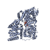
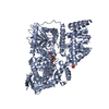
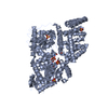
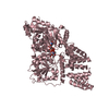
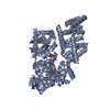
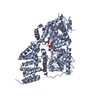
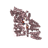
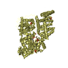
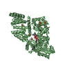
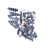
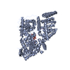
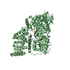
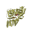
Chem-12V / UDP-N-acetylglucosamine--peptide N-acetylglucosaminyltransferase 110 kDa subunit / Keratin, type II cytoskeletal 7 Similarity search - Component
Biological species
Homo sapiens (human)
Method
X-RAY DIFFRACTION / SYNCHROTRON / Resolution: 3.2 Å
Protocol: SINGLE WAVELENGTH / Monochromatic (M) / Laue (L): M / Scattering type: x-ray
Radiation wavelength
Wavelength: 0.873 Å / Relative weight: 1
Reflection
Resolution: 3.2→25 Å / Num. obs: 105151 / % possible obs: 99 % / Redundancy: 1.7 % / Net I/σ(I): 1.7
-
Processing
Software
Name
Version
Classification
REFMAC
5.5.0088
refinement
SCALEPACK
datascaling
DENZO
datareduction
REFMAC
phasing
Refinement
Resolution: 3.2→25 Å / Cor.coef. Fo:Fc: 0.913 / Cor.coef. Fo:Fc free: 0.884 / SU B: 17.745 / SU ML: 0.29 / Cross valid method: THROUGHOUT / ESU R: 1.052 / ESU R Free: 0.365 / Stereochemistry target values: MAXIMUM LIKELIHOOD / Details: HYDROGENS HAVE BEEN ADDED IN THE RIDING POSITIONS
Rfactor
Num. reflection
% reflection
Selection details
Rfree
0.23893
1015
1 %
RANDOM
Rwork
0.2157
-
-
-
obs
0.21593
100304
99.01 %
-
Solvent computation
Ion probe radii: 0.8 Å / Shrinkage radii: 0.8 Å / VDW probe radii: 1.4 Å / Solvent model: MASK
 Movie
Movie Controller

 Yorodumi
 Open data
 Basic information
 Components
 Keywords
 TRANSFERASE / O-GlcNAc transferase Inverting GT-B Substrate complex
 Function and homology information
 Authors
 United Kingdom, 1items
  Citation
 Structure visualization
 Downloads & links
 Other downloads
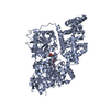


 PDBj
PDBj


 Assembly
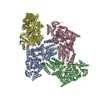


 Mass: 96.063 Da / Num. of mol.: 4 / Source method: obtained synthetically / Formula: SO4
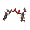
 Mass: 623.419 Da / Num. of mol.: 4 / Source method: obtained synthetically / Formula: C17H27N3O16P2S
 Sample preparation
 / Beamline: ID23-2 / Wavelength: 0.873 Å
 Processing