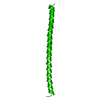
 Movie
Movie Controller


+ Open data

- Basic information
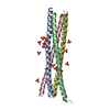
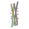
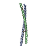
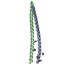
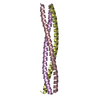
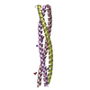
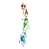
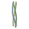
| Entry | Database: PDB / ID: 4w7y | ||||||
|---|---|---|---|---|---|---|---|
| Title | Dimeric BAP29 vDED with disulfide bonds in crystal contacts | ||||||
 Components Components | B-cell receptor-associated protein 29 | ||||||
 Keywords Keywords |  TRANSPORT PROTEIN / Coiled coil / nanomaterial TRANSPORT PROTEIN / Coiled coil / nanomaterial | ||||||
| Function / homology |  Function and homology information Function and homology informationprotein localization to endoplasmic reticulum exit site / endoplasmic reticulum to Golgi vesicle-mediated transport / intracellular protein transport / osteoblast differentiation / apoptotic process / endoplasmic reticulum membrane / membraneSimilarity search - Function | ||||||
| Biological species |  Homo sapiens (human) Homo sapiens (human) | ||||||
| Method | X-RAY DIFFRACTION / SYNCHROTRON / SAD / Resolution: 2.5 Å | ||||||
 Authors Authors | Quistgaard, E.M. | ||||||
 Citation Citation | Journal: Chem.Commun.(Camb.) / Year: 2014 Title: A disulfide polymerized protein crystal. Authors: Quistgaard, E.M. | ||||||
| History |
|
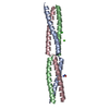
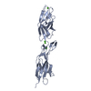
- Structure visualization
| Structure viewer | Molecule: MolmilJmol/JSmol |
|---|
- Downloads & links
-Download
| PDBx/mmCIF format | 4w7y.cif.gz | 63.2 KB | Display | PDBx/mmCIF format |
|---|---|---|---|---|
| PDB format | pdb4w7y.ent.gz | 51.3 KB | Display | PDB format |
| PDBx/mmJSON format | 4w7y.json.gz | Tree view | PDBx/mmJSON format | |
| Others |  Other downloads Other downloads |
-Validation report
| Arichive directory | https://data.pdbj.org/pub/pdb/validation_reports/w7/4w7yftp://data.pdbj.org/pub/pdb/validation_reports/w7/4w7y | HTTPS FTP |
|---|
-Related structure data

-Links
 PDBj
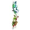
PDBj- Assembly
| Deposited unit | 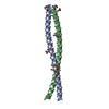
| ||||||||
|---|---|---|---|---|---|---|---|---|---|
| 1 |
| ||||||||
| Unit cell |
|
-Components
| #1: Protein | Mass: 7731.090 Da / Num. of mol.: 2 / Fragment: UNP residues 168-229 Source method: isolated from a genetically manipulated source Source: (gene. exp.) Homo sapiens (human) / Gene: BCAP29, BAP29 / Production host:  Escherichia coli (E. coli) / References: UniProt: Q9UHQ4 Escherichia coli (E. coli) / References: UniProt: Q9UHQ4#2: Chemical | 2-Methyl-2,4-pentanediol  Mass: 118.174 Da / Num. of mol.: 2 / Source method: obtained synthetically / Formula: C6H14O2 / Comment: precipitant*YM Mass: 118.174 Da / Num. of mol.: 2 / Source method: obtained synthetically / Formula: C6H14O2 / Comment: precipitant*YM#3: Chemical | ChemComp-ACT / | Acetate  Mass: 59.044 Da / Num. of mol.: 1 / Source method: obtained synthetically / Formula: C2H3O2 Mass: 59.044 Da / Num. of mol.: 1 / Source method: obtained synthetically / Formula: C2H3O2#4: Water | ChemComp-HOH / | Water Mass: 18.015 Da / Num. of mol.: 31 / Source method: isolated from a natural source / Formula: H2O Mass: 18.015 Da / Num. of mol.: 31 / Source method: isolated from a natural source / Formula: H2O |
|---|
-Experimental details
-Experiment
| Experiment | Method: X-RAY DIFFRACTION |
|---|
- Sample preparation
| Crystal | Density Matthews: 4.04 Å3/Da / Density % sol: 69.56 % |
|---|---|
| Crystal grow | Temperature: 292.15 K / Method: vapor diffusion / pH: 4.6 Details: Grown in 34% 2-methyl-2,4-pentanediol (MPD), 100 mM Na acetate pH 4.6 and 20 mM CaCl2 using SeMet labeled protein purified in 20 mM Tris pH 8, 150 mM NaCl and 2 mM tris(2-carboxyethyl) ...Details: Grown in 34% 2-methyl-2,4-pentanediol (MPD), 100 mM Na acetate pH 4.6 and 20 mM CaCl2 using SeMet labeled protein purified in 20 mM Tris pH 8, 150 mM NaCl and 2 mM tris(2-carboxyethyl)phosphine (TECEP) and concentrated to ~13 mg/mL. |
-Data collection
| Diffraction | Mean temperature: 100 K |
|---|---|
| Diffraction source | Source: SYNCHROTRON / Site: Diamond  / Beamline: I02 / Wavelength: 0.978 Å / Beamline: I02 / Wavelength: 0.978 Å |
| Detector | Type: ADSC QUANTUM 315 / Detector: CCD / Date: Oct 2, 2011 |
| Radiation | Protocol: SINGLE WAVELENGTH / Monochromatic (M) / Laue (L): M / Scattering type: x-ray |
| Radiation wavelength | Wavelength: 0.978 Å / Relative weight: 1 |
| Reflection | Resolution: 2.5→34.31 Å / Num. obs: 16720 / % possible obs: 99.9 % / Observed criterion σ(I): -3 / Redundancy: 6.4 % / Rsym value: 0.072 / Net I/σ(I): 17.09 |
| Reflection shell | Resolution: 2.5→2.64 Å / Redundancy: 6.3 % / Mean I/σ(I) obs: 2.93 / % possible all: 99.9 |
- Processing
| Software |
| |||||||||||||||||||||||||||||||||||||||||||||||||
|---|---|---|---|---|---|---|---|---|---|---|---|---|---|---|---|---|---|---|---|---|---|---|---|---|---|---|---|---|---|---|---|---|---|---|---|---|---|---|---|---|---|---|---|---|---|---|---|---|---|---|
| Refinement | Method to determine structure: SAD / Resolution: 2.5→34.315 Å / SU ML: 0.34 / Cross valid method: THROUGHOUT / σ(F): 1.47 / Phase error: 25.95 / Stereochemistry target values: ML
| |||||||||||||||||||||||||||||||||||||||||||||||||
| Solvent computation | Shrinkage radii: 0.9 Å / VDW probe radii: 1.11 Å / Solvent model: FLAT BULK SOLVENT MODEL | |||||||||||||||||||||||||||||||||||||||||||||||||
| Refinement step | Cycle: LAST / Resolution: 2.5→34.315 Å
| |||||||||||||||||||||||||||||||||||||||||||||||||
| Refine LS restraints |
| |||||||||||||||||||||||||||||||||||||||||||||||||
| LS refinement shell |
| |||||||||||||||||||||||||||||||||||||||||||||||||
| Refinement TLS params. | Method: refined / Origin x: 71.3112 Å / Origin y: -8.0539 Å / Origin z: 3.9062 Å
| |||||||||||||||||||||||||||||||||||||||||||||||||
| Refinement TLS group | Selection details: chain 'A' or chain 'B' |
