 Movie
Movie Controller


+ Open data

- Basic information
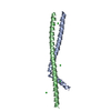
| Entry | Database: PDB / ID: 1t3j | ||||||
|---|---|---|---|---|---|---|---|
| Title | Mitofusin domain HR2 V686M/I708M mutant | ||||||
 Components Components | mitofusin 1 | ||||||
 Keywords Keywords |  MEMBRANE PROTEIN / coiled coil antiparallel / dimer MEMBRANE PROTEIN / coiled coil antiparallel / dimer | ||||||
| Function / homology |  Function and homology information Function and homology information: / mitochondrial fusion => GO:0008053 / : / : / outer mitochondrial membrane protein complex / PINK1-PRKN Mediated Mitophagy / positive regulation of mitochondrial fusion / mitochondrion localization / positive regulation of dendritic spine morphogenesis / Factors involved in megakaryocyte development and platelet production ...: / mitochondrial fusion => GO:0008053 / : / : / outer mitochondrial membrane protein complex / PINK1-PRKN Mediated Mitophagy / positive regulation of mitochondrial fusion / mitochondrion localization / positive regulation of dendritic spine morphogenesis / Factors involved in megakaryocyte development and platelet production / GTP metabolic process / positive regulation of mitochondrial membrane potential / mitochondrial fusion / intracellular distribution of mitochondria / negative regulation of mitochondrial fission / Hydrolases; Acting on acid anhydrides; Acting on GTP to facilitate cellular and subcellular movement / mitochondrial outer membrane / mitochondrial inner membrane / membrane => GO:0016020 / GTPase activity / GTP binding / mitochondrion / identical protein bindingSimilarity search - Function | ||||||
| Biological species |  Mus musculus (house mouse) Mus musculus (house mouse) | ||||||
| Method | X-RAY DIFFRACTION / SYNCHROTRON / MAD / Resolution: 2.5 Å | ||||||
 Authors Authors | Koshiba, T. / Detmer, S.A. / Kaiser, J.T. / Chen, H. / McCaffery, J.M. / Chan, D.C. | ||||||
 Citation Citation | Journal: Science / Year: 2004 Title: Structural basis of mitochondrial tethering by mitofusin complexes Authors: Koshiba, T. / Detmer, S.A. / Kaiser, J.T. / Chen, H. / McCaffery, J.M. / Chan, D.C. | ||||||
| History |
|
- Structure visualization
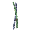
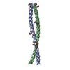
| Structure viewer | Molecule: MolmilJmol/JSmol |
|---|
- Downloads & links
-Download
| PDBx/mmCIF format | 1t3j.cif.gz | 25.4 KB | Display | PDBx/mmCIF format |
|---|---|---|---|---|
| PDB format | pdb1t3j.ent.gz | 17.8 KB | Display | PDB format |
| PDBx/mmJSON format | 1t3j.json.gz | Tree view | PDBx/mmJSON format | |
| Others |  Other downloads Other downloads |
-Validation report
| Arichive directory | https://data.pdbj.org/pub/pdb/validation_reports/t3/1t3jftp://data.pdbj.org/pub/pdb/validation_reports/t3/1t3j | HTTPS FTP |
|---|
-Related structure data
| Similar structure data |
|---|


-Links
 PDBj
PDBj


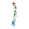
- Assembly
| Deposited unit | 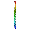
| ||||||||
|---|---|---|---|---|---|---|---|---|---|
| 1 | 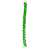
| ||||||||
| Unit cell |
| ||||||||
| Details | The second part of the dimer is generated by the twofold axis: -X+1/4, -Z+1/4, -Y+1/4 |
-Components
| #1: Protein | Mass: 11107.447 Da / Num. of mol.: 1 / Mutation: V686M, I708M Source method: isolated from a genetically manipulated source Source: (gene. exp.) Mus musculus (house mouse) / Gene: MFN1 / Plasmid: pET28a(+),pET28-MFN1-HR2 / Species (production host): Escherichia coli / Production host:  Escherichia coli BL21(DE3) (bacteria) / Strain (production host): BL21(DE3) / References: UniProt: Q811U4 Escherichia coli BL21(DE3) (bacteria) / Strain (production host): BL21(DE3) / References: UniProt: Q811U4 |
|---|---|
| #2: Water | ChemComp-HOH / Water Mass: 18.015 Da / Num. of mol.: 33 / Source method: isolated from a natural source / Formula: H2O Mass: 18.015 Da / Num. of mol.: 33 / Source method: isolated from a natural source / Formula: H2O |
-Experimental details
-Experiment
| Experiment | Method: X-RAY DIFFRACTION / Number of used crystals: 1 |
|---|
- Sample preparation
| Crystal | Density Matthews: 4.09 Å3/Da / Density % sol: 68 % |
|---|---|
| Crystal grow | Temperature: 290 K / Method: vapor diffusion, sitting drop / pH: 7.7 Details: Isopropanol, TRIS, Ammonium Acetate, PEG 200, pH 7.7, VAPOR DIFFUSION, SITTING DROP, temperature 295K, temperature 290K |
-Data collection
| Diffraction | Mean temperature: 100 K | |||||||||||||||
|---|---|---|---|---|---|---|---|---|---|---|---|---|---|---|---|---|
| Diffraction source | Source: SYNCHROTRON / Site: ALS  / Beamline: 8.2.1 / Wavelength: 0.9795, 0.9797, 0.9641, 0.9800 / Beamline: 8.2.1 / Wavelength: 0.9795, 0.9797, 0.9641, 0.9800 | |||||||||||||||
| Detector | Type: ADSC QUANTUM 210 / Detector: CCD / Date: Jan 22, 2004 | |||||||||||||||
| Radiation | Monochromator: Si(111) double crystal / Protocol: MAD / Monochromatic (M) / Laue (L): M / Scattering type: x-ray | |||||||||||||||
| Radiation wavelength |
| |||||||||||||||
| Reflection | Resolution: 2.5→27.52 Å / Num. all: 6772 / Num. obs: 6772 / % possible obs: 99.5 % / Observed criterion σ(F): 0 / Observed criterion σ(I): 0 / Redundancy: 10.5 % / Biso Wilson estimate: 44.1 Å2 / Limit h max: 65 / Limit h min: 5 / Limit k max: 46 / Limit k min: 5 / Limit l max: 37 / Limit l min: 0 / Observed criterion F max: 2907765.71 / Observed criterion F min: 14.9 / Rmerge(I) obs: 0.079 / Rsym value: 0.079 / Net I/σ(I): 25.2 | |||||||||||||||
| Reflection shell | Resolution: 2.5→2.54 Å / Rmerge(I) obs: 0.463 / Mean I/σ(I) obs: 5.6 / Num. unique all: 319 / Rsym value: 0.463 / % possible all: 99.2 |
- Processing
| Software |
| ||||||||||||||||||||||||||||||||||||||||||||||||||||||||||||||||||||||||||||||||||||||||||
|---|---|---|---|---|---|---|---|---|---|---|---|---|---|---|---|---|---|---|---|---|---|---|---|---|---|---|---|---|---|---|---|---|---|---|---|---|---|---|---|---|---|---|---|---|---|---|---|---|---|---|---|---|---|---|---|---|---|---|---|---|---|---|---|---|---|---|---|---|---|---|---|---|---|---|---|---|---|---|---|---|---|---|---|---|---|---|---|---|---|---|---|
| Refinement | Method to determine structure: MAD / Resolution: 2.5→14.86 Å / Rfactor Rfree error: 0.011 / Occupancy max: 1 / Occupancy min: 1 / Isotropic thermal model: isotropic / Cross valid method: THROUGHOUT / σ(F): 0 / Stereochemistry target values: Engh & Huber
| ||||||||||||||||||||||||||||||||||||||||||||||||||||||||||||||||||||||||||||||||||||||||||
| Solvent computation | Solvent model: bulk solvent / Bsol: 52.5258 Å2 / ksol: 0.388799 e/Å3 | ||||||||||||||||||||||||||||||||||||||||||||||||||||||||||||||||||||||||||||||||||||||||||
| Displacement parameters | Biso max: 99.67 Å2 / Biso mean: 52.81 Å2 / Biso min: 26.39 Å2 | ||||||||||||||||||||||||||||||||||||||||||||||||||||||||||||||||||||||||||||||||||||||||||
| Refine analyze |
| ||||||||||||||||||||||||||||||||||||||||||||||||||||||||||||||||||||||||||||||||||||||||||
| Refinement step | Cycle: LAST / Resolution: 2.5→14.86 Å
| ||||||||||||||||||||||||||||||||||||||||||||||||||||||||||||||||||||||||||||||||||||||||||
| Refine LS restraints |
| ||||||||||||||||||||||||||||||||||||||||||||||||||||||||||||||||||||||||||||||||||||||||||
| LS refinement shell | Refine-ID: X-RAY DIFFRACTION / Total num. of bins used: 8
| ||||||||||||||||||||||||||||||||||||||||||||||||||||||||||||||||||||||||||||||||||||||||||
| Xplor file |
|
