 Movie
Movie Controller

[English] 日本語
 Yorodumi
- PDB-4uz3: Crystal structure of the N-terminal LysM domains from the putativ... -
+ Open data

- Basic information
| Entry | Database: PDB / ID: 4uz3 | |||||||||
|---|---|---|---|---|---|---|---|---|---|---|
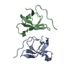
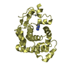
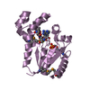
| Title | Crystal structure of the N-terminal LysM domains from the putative NlpC/P60 D,L endopeptidase from T. thermophilus bound to N-acetyl-chitohexaose | |||||||||
 Components Components | CELL WALL-BINDING ENDOPEPTIDASE-RELATED PROTEIN | |||||||||
 Keywords Keywords |  HYDROLASE HYDROLASE | |||||||||
| Function / homology |  Function and homology information Function and homology informationMembrane-bound Lytic Murein Transglycosylase D; Chain A / LysM domain / NlpC/P60 domain profile. / Endopeptidase, NLPC/P60 domain / NlpC/P60 family / Lysin motif / LysM domain superfamily / LysM domain / LysM domain profile. / LysM domain ...Membrane-bound Lytic Murein Transglycosylase D; Chain A / LysM domain / NlpC/P60 domain profile. / Endopeptidase, NLPC/P60 domain / NlpC/P60 family / Lysin motif / LysM domain superfamily / LysM domain / LysM domain profile. / LysM domain / Papain-like cysteine peptidase superfamily / Roll / Alpha BetaSimilarity search - Domain/homology | |||||||||
| Biological species |   THERMUS THERMOPHILUS (bacteria) THERMUS THERMOPHILUS (bacteria) | |||||||||
| Method | X-RAY DIFFRACTION / SYNCHROTRON / MOLECULAR REPLACEMENT / Resolution: 1.75 Å | |||||||||
 Authors Authors | Wong, J.E.M.M. / Blaise, M. | |||||||||
 Citation Citation | Journal: Acta Crystallogr.,Sect.D / Year: 2015 Title: An Intermolecular Binding Mechanism Involving Multiple Lysm Domains Mediates Carbohydrate Recognition by an Endopeptidase. Authors: Wong, J.E.M.M. / Midtgaard, S.R. / Gysel, K. / Thygesen, M.B. / Sorensen, K.K. / Jensen, K.J. / Stougaard, J. / Thirup, S. / Blaise, M. | |||||||||
| History |
|
- Structure visualization
| Structure viewer | Molecule: MolmilJmol/JSmol |
|---|
- Downloads & links
-Download
| PDBx/mmCIF format | 4uz3.cif.gz | 127.3 KB | Display | PDBx/mmCIF format |
|---|---|---|---|---|
| PDB format | pdb4uz3.ent.gz | 102 KB | Display | PDB format |
| PDBx/mmJSON format | 4uz3.json.gz | Tree view | PDBx/mmJSON format | |
| Others |  Other downloads Other downloads |
-Validation report
| Arichive directory | https://data.pdbj.org/pub/pdb/validation_reports/uz/4uz3ftp://data.pdbj.org/pub/pdb/validation_reports/uz/4uz3 | HTTPS FTP |
|---|
-Related structure data


-Links
 PDBj
PDBj


- Assembly
| Deposited unit | 
| ||||||||
|---|---|---|---|---|---|---|---|---|---|
| 1 | 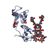
| ||||||||
| 2 | 
| ||||||||
| 3 | 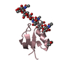
| ||||||||
| Unit cell |
| ||||||||
| Components on special symmetry positions |
|
-Components
-Protein , 1 types, 3 molecules ABC
| #1: Protein | Mass: 10971.500 Da / Num. of mol.: 3 / Fragment: LYSM DOMAIN, RESIDUES 16-114 Source method: isolated from a genetically manipulated source Source: (gene. exp.) THERMUS THERMOPHILUS (bacteria) / Strain: HB8 / Production host: ESCHERICHIA COLI (E. coli) / Strain (production host): BL21(DE3) / Variant (production host): ROSETTA2 / References: UniProt: Q5SLM7 |
|---|
-Sugars , 4 types, 4 molecules 
| #2: Polysaccharide | 2-acetamido-2-deoxy-beta-D-glucopyranose-(1-4)-2-acetamido-2-deoxy-beta-D-glucopyranose-(1-4)-2- ...2-acetamido-2-deoxy-beta-D-glucopyranose-(1-4)-2-acetamido-2-deoxy-beta-D-glucopyranose-(1-4)-2-acetamido-2-deoxy-beta-D-glucopyranose-(1-4)-2-acetamido-2-deoxy-beta-D-glucopyranose-(1-4)-2-acetamido-2-deoxy-beta-D-glucopyranose-(1-4)-2-acetamido-2-deoxy-alpha-D-glucopyranose / Mass: 1237.172 Da / Num. of mol.: 1 Source method: isolated from a genetically manipulated source |
|---|---|
| #3: Polysaccharide | 2-acetamido-2-deoxy-beta-D-glucopyranose-(1-4)-2-acetamido-2-deoxy-beta-D-glucopyranose-(1-4)-2- ...2-acetamido-2-deoxy-beta-D-glucopyranose-(1-4)-2-acetamido-2-deoxy-beta-D-glucopyranose-(1-4)-2-acetamido-2-deoxy-beta-D-glucopyranose-(1-4)-2-acetamido-2-deoxy-beta-D-glucopyranose-(1-4)-2-acetamido-2-deoxy-beta-D-glucopyranose-(1-4)-2-acetamido-2-deoxy-beta-D-glucopyranose / Mass: 1237.172 Da / Num. of mol.: 1 Source method: isolated from a genetically manipulated source |
| #4: Polysaccharide | 2-acetamido-2-deoxy-beta-D-glucopyranose-(1-4)-2-acetamido-2-deoxy-beta-D-glucopyranose-(1-4)-2- ...2-acetamido-2-deoxy-beta-D-glucopyranose-(1-4)-2-acetamido-2-deoxy-beta-D-glucopyranose-(1-4)-2-acetamido-2-deoxy-beta-D-glucopyranose-(1-4)-2-acetamido-2-deoxy-beta-D-glucopyranose-(1-4)-2-acetamido-2-deoxy-beta-D-glucopyranose-(1-4)-2-acetamido-2-deoxy-beta-D-glucopyranose / Mass: 1237.172 Da / Num. of mol.: 1 Source method: isolated from a genetically manipulated source |
| #5: Sugar | ChemComp-NDG / N-Acetylglucosamine Type: D-saccharide, alpha linking / Mass: 221.208 Da / Num. of mol.: 1 Type: D-saccharide, alpha linking / Mass: 221.208 Da / Num. of mol.: 1Source method: isolated from a genetically manipulated source Formula: C8H15NO6 |
-Non-polymers , 3 types, 398 molecules 


| #6: Chemical | Sulfate Mass: 96.063 Da / Num. of mol.: 3 / Source method: obtained synthetically / Formula: SO4 Mass: 96.063 Da / Num. of mol.: 3 / Source method: obtained synthetically / Formula: SO4#7: Chemical | 1,4-Dioxane Mass: 88.105 Da / Num. of mol.: 2 / Source method: obtained synthetically / Formula: C4H8O2 Mass: 88.105 Da / Num. of mol.: 2 / Source method: obtained synthetically / Formula: C4H8O2#8: Water | ChemComp-HOH / | WaterMass: 18.015 Da / Num. of mol.: 393 / Source method: isolated from a natural source / Formula: H2O |
|---|
-Experimental details
-Experiment
| Experiment | Method: X-RAY DIFFRACTION / Number of used crystals: 1 |
|---|
- Sample preparation
| Crystal | Density Matthews: 2.97 Å3/Da / Density % sol: 58.61 % / Description: NONE |
|---|---|
| Crystal grow | Details: 0.1 M MES PH6.5, 1.6 M AMMONIUM SULFATE, 5% DIOXANE |
-Data collection
| Diffraction | Mean temperature: 100 K |
|---|---|
| Diffraction source | Source: SYNCHROTRON / Site: MAX II  / Beamline: I911-2 / Wavelength: 1 / Beamline: I911-2 / Wavelength: 1 |
| Detector | Type: MARRESEARCH / Detector: CCD / Date: Nov 27, 2013 |
| Radiation | Protocol: SINGLE WAVELENGTH / Monochromatic (M) / Laue (L): M / Scattering type: x-ray |
| Radiation wavelength | Wavelength: 1 Å / Relative weight: 1 |
| Reflection | Resolution: 1.75→20 Å / Num. obs: 39631 / % possible obs: 99.9 % / Observed criterion σ(I): 2 / Redundancy: 12.3 % / Biso Wilson estimate: 14.78 Å2 / Rmerge(I) obs: 0.01 / Net I/σ(I): 20.3 |
| Reflection shell | Resolution: 1.75→1.8 Å / Redundancy: 12.3 % / Rmerge(I) obs: 0.87 / Mean I/σ(I) obs: 3.3 / % possible all: 100 |
- Processing
| Software |
| |||||||||||||||||||||||||||||||||||||||||||||||||||||||||||||||||||||||||||||||||||||||||||||||||||||||||
|---|---|---|---|---|---|---|---|---|---|---|---|---|---|---|---|---|---|---|---|---|---|---|---|---|---|---|---|---|---|---|---|---|---|---|---|---|---|---|---|---|---|---|---|---|---|---|---|---|---|---|---|---|---|---|---|---|---|---|---|---|---|---|---|---|---|---|---|---|---|---|---|---|---|---|---|---|---|---|---|---|---|---|---|---|---|---|---|---|---|---|---|---|---|---|---|---|---|---|---|---|---|---|---|---|---|---|
| Refinement | Method to determine structure: MOLECULAR REPLACEMENT / Resolution: 1.75→19.589 Å / SU ML: 0.16 / σ(F): 1.37 / Phase error: 17.3 / Stereochemistry target values: ML
| |||||||||||||||||||||||||||||||||||||||||||||||||||||||||||||||||||||||||||||||||||||||||||||||||||||||||
| Solvent computation | Shrinkage radii: 0.9 Å / VDW probe radii: 1.11 Å / Solvent model: FLAT BULK SOLVENT MODEL | |||||||||||||||||||||||||||||||||||||||||||||||||||||||||||||||||||||||||||||||||||||||||||||||||||||||||
| Displacement parameters | Biso mean: 18.7 Å2 | |||||||||||||||||||||||||||||||||||||||||||||||||||||||||||||||||||||||||||||||||||||||||||||||||||||||||
| Refinement step | Cycle: LAST / Resolution: 1.75→19.589 Å
| |||||||||||||||||||||||||||||||||||||||||||||||||||||||||||||||||||||||||||||||||||||||||||||||||||||||||
| Refine LS restraints |
| |||||||||||||||||||||||||||||||||||||||||||||||||||||||||||||||||||||||||||||||||||||||||||||||||||||||||
| LS refinement shell |
|
