 Movie
Movie Controller


+ Open data

- Basic information
| Entry | Database: PDB / ID: 6jjz | ||||||
|---|---|---|---|---|---|---|---|
| Title | Crystal Structure of MAGI2 and Dendrin complex | ||||||
 Components Components |
| ||||||
 Keywords Keywords |  SIGNALING PROTEIN / WW tandem / PY tandem / MAGI2 / Dendrin SIGNALING PROTEIN / WW tandem / PY tandem / MAGI2 / Dendrin | ||||||
| Function / homology |  Function and homology information Function and homology informationstructural constituent of postsynaptic specialization / extrinsic component of postsynaptic membrane / type II activin receptor binding / neuroligin clustering involved in postsynaptic membrane assembly / podocyte development / positive regulation of synaptic vesicle clustering / slit diaphragm / beta-1 adrenergic receptor binding / structural constituent of postsynaptic density / nerve growth factor signaling pathway ...structural constituent of postsynaptic specialization / extrinsic component of postsynaptic membrane / type II activin receptor binding / neuroligin clustering involved in postsynaptic membrane assembly / podocyte development / positive regulation of synaptic vesicle clustering / slit diaphragm / beta-1 adrenergic receptor binding / structural constituent of postsynaptic density / nerve growth factor signaling pathway / activin receptor binding / clathrin-dependent endocytosis / negative regulation of activin receptor signaling pathway / SMAD protein signal transduction / dendritic spine membrane / regulation of neurotransmitter receptor localization to postsynaptic specialization membrane / regulation of synaptic membrane adhesion / ciliary base / SMAD binding / receptor clustering / kinesin binding / positive regulation of phosphoprotein phosphatase activity / positive regulation of receptor internalization / photoreceptor outer segment / GABA-ergic synapse / bicellular tight junction / negative regulation of phosphatidylinositol 3-kinase/protein kinase B signal transduction / phosphatase binding / centriole / photoreceptor inner segment / negative regulation of cell migration / cellular response to nerve growth factor stimulus / cell projection / positive regulation of neuron projection development / cell-cell junction / signaling receptor complex adaptor activity / late endosome / postsynaptic membrane / perikaryon / postsynaptic density / DNA-binding transcription factor activity, RNA polymerase II-specific / RNA polymerase II cis-regulatory region sequence-specific DNA binding / negative regulation of cell population proliferation / centrosome / glutamatergic synapse / synapse / dendrite / protein-containing complex binding / endoplasmic reticulum membrane / perinuclear region of cytoplasm / signal transduction / positive regulation of transcription by RNA polymerase II / protein-containing complex / nucleus / plasma membrane / cytoplasmSimilarity search - Function | ||||||
| Biological species |  Mus musculus (house mouse) Mus musculus (house mouse) | ||||||
| Method | X-RAY DIFFRACTION / SYNCHROTRON / MOLECULAR REPLACEMENT / Resolution: 1.65 Å | ||||||
 Authors Authors | Lin, Z. / Zhang, H. / Yang, Z. / Ji, Z. / Zhang, M. / Zhu, J. | ||||||
 Citation Citation | Journal: Elife / Year: 2019 Title: Decoding WW domain tandem-mediated target recognitions in tissue growth and cell polarity. Authors: Lin, Z. / Yang, Z. / Xie, R. / Ji, Z. / Guan, K. / Zhang, M. | ||||||
| History |
|
- Structure visualization
| Structure viewer | Molecule: MolmilJmol/JSmol |
|---|
- Downloads & links
-Download
| PDBx/mmCIF format | 6jjz.cif.gz | 106.4 KB | Display | PDBx/mmCIF format |
|---|---|---|---|---|
| PDB format | pdb6jjz.ent.gz | 80.2 KB | Display | PDB format |
| PDBx/mmJSON format | 6jjz.json.gz | Tree view | PDBx/mmJSON format | |
| Others |  Other downloads Other downloads |
-Validation report
| Arichive directory | https://data.pdbj.org/pub/pdb/validation_reports/jj/6jjzftp://data.pdbj.org/pub/pdb/validation_reports/jj/6jjz | HTTPS FTP |
|---|
-Related structure data
| Related structure data | 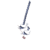 6j68C 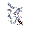 6jjwC 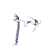 6jjxC 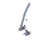 6jjyC 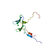 6jk0C 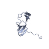 6jk1C  2ysdS S: Starting model for refinement C: citing same article ( |
|---|---|
| Similar structure data |


-Links
 PDBj
PDBj
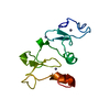
- Assembly
| Deposited unit | 
| ||||||||
|---|---|---|---|---|---|---|---|---|---|
| 1 | 
| ||||||||
| 2 | 
| ||||||||
| Unit cell |
|
-Components
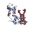
| #1: Protein | / Activin receptor-interacting protein 1 / Acvrip1 / Atrophin-1-interacting protein 1 / AIP-1 / ...Activin receptor-interacting protein 1 / Acvrip1 / Atrophin-1-interacting protein 1 / AIP-1 / Membrane-associated guanylate kinase inverted 2 / MAGI-2 Mass: 12089.212 Da / Num. of mol.: 2 Source method: isolated from a genetically manipulated source Source: (gene. exp.) Mus musculus (house mouse) / Gene: Magi2, Acvrinp1, Aip1, Arip1 / Production host:  Escherichia coli (E. coli) / References: UniProt: Q9WVQ1 Escherichia coli (E. coli) / References: UniProt: Q9WVQ1#2: Protein/peptide | Mass: 1843.966 Da / Num. of mol.: 2 Source method: isolated from a genetically manipulated source Source: (gene. exp.) Mus musculus (house mouse) / Gene: Ddn, Gm748, Kiaa0749 / Production host: Escherichia coli (E. coli) / References: UniProt: Q80TS7#3: Chemical | Acetate  Mass: 59.044 Da / Num. of mol.: 2 / Source method: obtained synthetically / Formula: C2H3O2 Mass: 59.044 Da / Num. of mol.: 2 / Source method: obtained synthetically / Formula: C2H3O2#4: Chemical | ChemComp-SO4 / | Sulfate  Mass: 96.063 Da / Num. of mol.: 1 / Source method: obtained synthetically / Formula: SO4 Mass: 96.063 Da / Num. of mol.: 1 / Source method: obtained synthetically / Formula: SO4#5: Water | ChemComp-HOH / | Water Mass: 18.015 Da / Num. of mol.: 144 / Source method: isolated from a natural source / Formula: H2O Mass: 18.015 Da / Num. of mol.: 144 / Source method: isolated from a natural source / Formula: H2O |
|---|
-Experimental details
-Experiment
| Experiment | Method: X-RAY DIFFRACTION / Number of used crystals: 1 |
|---|
- Sample preparation
| Crystal | Density Matthews: 2.13 Å3/Da / Density % sol: 42.17 % |
|---|---|
| Crystal grow | Temperature: 289 K / Method: vapor diffusion / pH: 4.6 Details: 2.0-2.5M ammonium sulfate, 100mM sodium acetate (pH 4.6) |
-Data collection
| Diffraction | Mean temperature: 100 K / Serial crystal experiment: N |
|---|---|
| Diffraction source | Source: SYNCHROTRON / Site: SSRF  / Beamline: BL17U1 / Wavelength: 0.97915 Å / Beamline: BL17U1 / Wavelength: 0.97915 Å |
| Detector | Type: ADSC QUANTUM 315r / Detector: CCD / Date: Mar 20, 2016 |
| Radiation | Protocol: SINGLE WAVELENGTH / Monochromatic (M) / Laue (L): M / Scattering type: x-ray |
| Radiation wavelength | Wavelength: 0.97915 Å / Relative weight: 1 |
| Reflection | Resolution: 1.65→50 Å / Num. obs: 29381 / % possible obs: 99.8 % / Redundancy: 6.9 % / Rmerge(I) obs: 0.047 / Net I/σ(I): 44 |
| Reflection shell | Resolution: 1.65→1.68 Å / Redundancy: 6.8 % / Rmerge(I) obs: 0.856 / Mean I/σ(I) obs: 2.5 / Num. unique obs: 1465 / % possible all: 99.9 |
- Processing
| Software |
| |||||||||||||||||||||||||||||||||||||||||||||||||||||||||||||||||||||||||||||
|---|---|---|---|---|---|---|---|---|---|---|---|---|---|---|---|---|---|---|---|---|---|---|---|---|---|---|---|---|---|---|---|---|---|---|---|---|---|---|---|---|---|---|---|---|---|---|---|---|---|---|---|---|---|---|---|---|---|---|---|---|---|---|---|---|---|---|---|---|---|---|---|---|---|---|---|---|---|---|
| Refinement | Method to determine structure: MOLECULAR REPLACEMENT Starting model: 2YSD Resolution: 1.65→27.917 Å / SU ML: 0.2 / Cross valid method: FREE R-VALUE / σ(F): 1.89 / Phase error: 27.42
| |||||||||||||||||||||||||||||||||||||||||||||||||||||||||||||||||||||||||||||
| Solvent computation | Shrinkage radii: 0.9 Å / VDW probe radii: 1.11 Å | |||||||||||||||||||||||||||||||||||||||||||||||||||||||||||||||||||||||||||||
| Refinement step | Cycle: LAST / Resolution: 1.65→27.917 Å
| |||||||||||||||||||||||||||||||||||||||||||||||||||||||||||||||||||||||||||||
| Refine LS restraints |
| |||||||||||||||||||||||||||||||||||||||||||||||||||||||||||||||||||||||||||||
| LS refinement shell |
| |||||||||||||||||||||||||||||||||||||||||||||||||||||||||||||||||||||||||||||
| Refinement TLS params. | Method: refined / Origin x: -9.2337 Å / Origin y: -5.2763 Å / Origin z: 13.4028 Å
| |||||||||||||||||||||||||||||||||||||||||||||||||||||||||||||||||||||||||||||
| Refinement TLS group | Selection details: all |
