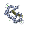
SEQUENCE The cysteines at residue positions 47, 155, 171 and 184 were converted to serines by site- ...SEQUENCE The cysteines at residue positions 47, 155, 171 and 184 were converted to serines by site-directed mutagenesis in the construct. These substitutions do not correspond to known mutations in the Deltex protein, but were made to improve the solubility and long-term stability of the polypeptide.
Method to determine structure: MAD / Resolution: 2.15→40.8 Å / σ(F): 0 / Stereochemistry target values: Engh & Huber / Details: The sf file of this entry contains Friedel pairs.
Rfactor
Num. reflection
% reflection
Selection details
Rfree
0.247
1300
4.6 %
RANDOM
Rwork
0.23
-
-
-
all
-
28099
-
-
obs
-
26923
95.8 %
-
Solvent computation
Bsol: 56.987 Å2
Displacement parameters
Biso mean: 55.148 Å2
Baniso -1
Baniso -2
Baniso -3
1-
-0.803 Å2
0 Å2
0 Å2
2-
-
-0.803 Å2
0 Å2
3-
-
-
1.607 Å2
Refinement step
Cycle: LAST / Resolution: 2.15→40.8 Å
Protein
Nucleic acid
Ligand
Solvent
Total
Num. atoms
1286
0
0
54
1340
LS refinement shell
Resolution: 2.15→2.28 Å / Rfactor Rfree error: 0.023 / Total num. of bins used: 6
Rfactor
Num. reflection
% reflection
Rfree
0.335
204
4.9 %
Rwork
0.312
3957
-
obs
-
-
89.6 %
Xplor file
Refine-ID
Serial no
Param file
X-RAY DIFFRACTION
1
CNS_TOPPAR:protein_rep.param
X-RAY DIFFRACTION
2
CNS_TOPPAR:water_rep.param
+
About Yorodumi
-
News
-
Feb 9, 2022. New format data for meta-information of EMDB entries
New format data for meta-information of EMDB entries
Version 3 of the EMDB header file is now the official format.
The previous official version 1.9 will be removed from the archive.
In the structure databanks used in Yorodumi, some data are registered as the other names, "COVID-19 virus" and "2019-nCoV". Here are the details of the virus and the list of structure data.
Jan 31, 2019. EMDB accession codes are about to change! (news from PDBe EMDB page)
EMDB accession codes are about to change! (news from PDBe EMDB page)
The allocation of 4 digits for EMDB accession codes will soon come to an end. Whilst these codes will remain in use, new EMDB accession codes will include an additional digit and will expand incrementally as the available range of codes is exhausted. The current 4-digit format prefixed with “EMD-” (i.e. EMD-XXXX) will advance to a 5-digit format (i.e. EMD-XXXXX), and so on. It is currently estimated that the 4-digit codes will be depleted around Spring 2019, at which point the 5-digit format will come into force.
The EM Navigator/Yorodumi systems omit the EMD- prefix.
Related info.:Q: What is EMD? / ID/Accession-code notation in Yorodumi/EM Navigator
Yorodumi is a browser for structure data from EMDB, PDB, SASBDB, etc.
This page is also the successor to EM Navigator detail page, and also detail information page/front-end page for Omokage search.
The word "yorodu" (or yorozu) is an old Japanese word meaning "ten thousand". "mi" (miru) is to see.
Related info.:EMDB / PDB / SASBDB / Comparison of 3 databanks / Yorodumi Search / Aug 31, 2016. New EM Navigator & Yorodumi / Yorodumi Papers / Jmol/JSmol / Function and homology information / Changes in new EM Navigator and Yorodumi
 Movie
Movie Controller


 Open data
 Basic information
 Components
 Keywords
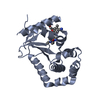
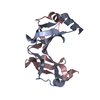
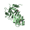
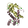
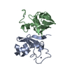
 WWE domain
 Function and homology information
 Authors
 Citation
 Structure visualization
 Downloads & links
 Other downloads
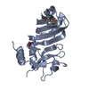


 PDBj
PDBj

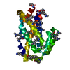
 Assembly

 Mass: 18.015 Da / Num. of mol.: 54 / Source method: isolated from a natural source / Formula: H2O
 Sample preparation
 / Beamline: X4A / Wavelength: 0.9795, 0.9793, 0.9724
 Processing