 Movie
Movie Controller


+ Open data

- Basic information
| Entry | Database: PDB / ID: 6wn7 | ||||||
|---|---|---|---|---|---|---|---|
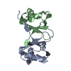
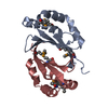
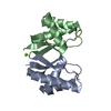
| Title | Homo sapiens S100A5 | ||||||
 Components Components | Protein S100-A5 | ||||||
 Keywords Keywords | METAL BINDING PROTEIN / S100 /  calcium / HA5 / EF-Hand calcium / HA5 / EF-Hand | ||||||
| Function / homology |  Function and homology information Function and homology informationcalcium-dependent protein binding / copper ion binding / neuronal cell body / calcium ion binding / protein homodimerization activity / zinc ion binding / nucleusSimilarity search - Function | ||||||
| Biological species |  Homo sapiens (human) Homo sapiens (human) | ||||||
| Method | X-RAY DIFFRACTION / SYNCHROTRON / MOLECULAR REPLACEMENT / Resolution: 1.25 Å | ||||||
 Authors Authors | Perkins, A. / Harms, M.J. / Wong, C.E. / Wheeler, L.C. | ||||||
 Citation Citation | Journal: Protein Sci. / Year: 2020 Title: Learning peptide recognition rules for a low-specificity protein. Authors: Wheeler, L.C. / Perkins, A. / Wong, C.E. / Harms, M.J. | ||||||
| History |
|
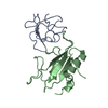
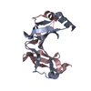
- Structure visualization
| Structure viewer | Molecule: MolmilJmol/JSmol |
|---|
- Downloads & links
-Download
| PDBx/mmCIF format | 6wn7.cif.gz | 351.7 KB | Display | PDBx/mmCIF format |
|---|---|---|---|---|
| PDB format | pdb6wn7.ent.gz | 258.3 KB | Display | PDB format |
| PDBx/mmJSON format | 6wn7.json.gz | Tree view | PDBx/mmJSON format | |
| Others |  Other downloads Other downloads |
-Validation report
| Arichive directory | https://data.pdbj.org/pub/pdb/validation_reports/wn/6wn7ftp://data.pdbj.org/pub/pdb/validation_reports/wn/6wn7 | HTTPS FTP |
|---|
-Related structure data
| Related structure data | 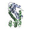 4dirS S: Starting model for refinement |
|---|---|
| Similar structure data |


-Links
 PDBj
PDBj- Assembly
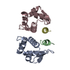
| Deposited unit | 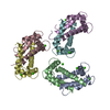
| ||||||||||||
|---|---|---|---|---|---|---|---|---|---|---|---|---|---|
| 1 | 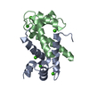
| ||||||||||||
| 2 | 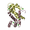
| ||||||||||||
| 3 | 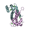
| ||||||||||||
| Unit cell |
|
-Components
| #1: Protein | Mass: 10999.547 Da / Num. of mol.: 6 / Mutation: C43S, C80S Source method: isolated from a genetically manipulated source Source: (gene. exp.) Homo sapiens (human) / Gene: S100A5, S100D / Production host:  Escherichia coli (E. coli) / References: UniProt: P33763 Escherichia coli (E. coli) / References: UniProt: P33763#2: Chemical | ChemComp-CA /   Mass: 40.078 Da / Num. of mol.: 18 / Source method: isolated from a natural source / Formula: Ca Mass: 40.078 Da / Num. of mol.: 18 / Source method: isolated from a natural source / Formula: Ca#3: Water | ChemComp-HOH / | Water Mass: 18.015 Da / Num. of mol.: 399 / Source method: isolated from a natural source / Formula: H2O Mass: 18.015 Da / Num. of mol.: 399 / Source method: isolated from a natural source / Formula: H2OHas ligand of interest | N | |
|---|
-Experimental details
-Experiment
| Experiment | Method: X-RAY DIFFRACTION / Number of used crystals: 1 |
|---|
- Sample preparation
| Crystal | Density Matthews: 2.26 Å3/Da / Density % sol: 45.49 % |
|---|---|
| Crystal grow | Temperature: 277 K / Method: vapor diffusion, hanging drop Details: 0.2 M ammonium sulfate, 20% w/v PEG8000, cryoprotectant: 25% PEG1500 |
-Data collection
| Diffraction | Mean temperature: 100 K / Serial crystal experiment: N |
|---|---|
| Diffraction source | Source: SYNCHROTRON / Site: ALS  / Beamline: 5.0.3 / Wavelength: 1 Å / Beamline: 5.0.3 / Wavelength: 1 Å |
| Detector | Type: ADSC QUANTUM 315r / Detector: CCD / Date: Mar 25, 2018 |
| Radiation | Monochromator: Si(220) / Protocol: SINGLE WAVELENGTH / Monochromatic (M) / Laue (L): M / Scattering type: x-ray |
| Radiation wavelength | Wavelength: 1 Å / Relative weight: 1 |
| Reflection | Resolution: 1.25→51.98 Å / Num. obs: 151437 / % possible obs: 99.8 % / Redundancy: 10.6 % / Biso Wilson estimate: 14.87 Å2 / CC1/2: 1 / Net I/σ(I): 16.2 |
| Reflection shell | Resolution: 1.25→1.32 Å / Redundancy: 8.4 % / Num. unique obs: 4561 / CC1/2: 0.48 / % possible all: 94.6 |
- Processing
| Software |
| |||||||||||||||||||||||||||||||||||||||||||||||||||||||||||||||||||||||||||||||||||||||||||||||||||||||||||||||||||||||||||||||||||||||||||||||||||
|---|---|---|---|---|---|---|---|---|---|---|---|---|---|---|---|---|---|---|---|---|---|---|---|---|---|---|---|---|---|---|---|---|---|---|---|---|---|---|---|---|---|---|---|---|---|---|---|---|---|---|---|---|---|---|---|---|---|---|---|---|---|---|---|---|---|---|---|---|---|---|---|---|---|---|---|---|---|---|---|---|---|---|---|---|---|---|---|---|---|---|---|---|---|---|---|---|---|---|---|---|---|---|---|---|---|---|---|---|---|---|---|---|---|---|---|---|---|---|---|---|---|---|---|---|---|---|---|---|---|---|---|---|---|---|---|---|---|---|---|---|---|---|---|---|---|---|---|---|
| Refinement | Method to determine structure: MOLECULAR REPLACEMENT Starting model: PDB entry 4DIR Resolution: 1.25→26 Å / Cross valid method: FREE R-VALUE / σ(F): 0.01 / Phase error: 37.2278 Stereochemistry target values: GeoStd + Monomer Library + CDL v1.2 Details: Data is twinned, as was refined with a twin law of -k, -h, -l and twin fraction of 0.44.
| |||||||||||||||||||||||||||||||||||||||||||||||||||||||||||||||||||||||||||||||||||||||||||||||||||||||||||||||||||||||||||||||||||||||||||||||||||
| Solvent computation | Shrinkage radii: 0.9 Å / VDW probe radii: 1.11 Å / Solvent model: FLAT BULK SOLVENT MODEL | |||||||||||||||||||||||||||||||||||||||||||||||||||||||||||||||||||||||||||||||||||||||||||||||||||||||||||||||||||||||||||||||||||||||||||||||||||
| Displacement parameters | Biso mean: 16.55 Å2 | |||||||||||||||||||||||||||||||||||||||||||||||||||||||||||||||||||||||||||||||||||||||||||||||||||||||||||||||||||||||||||||||||||||||||||||||||||
| Refinement step | Cycle: LAST / Resolution: 1.25→26 Å
| |||||||||||||||||||||||||||||||||||||||||||||||||||||||||||||||||||||||||||||||||||||||||||||||||||||||||||||||||||||||||||||||||||||||||||||||||||
| Refine LS restraints |
| |||||||||||||||||||||||||||||||||||||||||||||||||||||||||||||||||||||||||||||||||||||||||||||||||||||||||||||||||||||||||||||||||||||||||||||||||||
| LS refinement shell |
| |||||||||||||||||||||||||||||||||||||||||||||||||||||||||||||||||||||||||||||||||||||||||||||||||||||||||||||||||||||||||||||||||||||||||||||||||||
| Refinement TLS params. | Method: refined / Origin x: 20.4939936122 Å / Origin y: 3.41248205829 Å / Origin z: -7.00332290166 Å
| |||||||||||||||||||||||||||||||||||||||||||||||||||||||||||||||||||||||||||||||||||||||||||||||||||||||||||||||||||||||||||||||||||||||||||||||||||
| Refinement TLS group | Selection details: all |
