 Movie
Movie Controller

[English] 日本語
 Yorodumi
- PDB-4tve: Structure Of the First Two Thioredoxin Domains of Naumovozyma dai... -
+ Open data

- Basic information
| Entry | Database: PDB / ID: 4tve | |||||||||
|---|---|---|---|---|---|---|---|---|---|---|
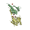
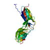
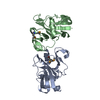
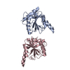
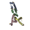
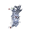
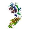
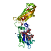
| Title | Structure Of the First Two Thioredoxin Domains of Naumovozyma dairenensis Eps1p | |||||||||
 Components Components | Naumovozyma dairenensis Eps1p | |||||||||
 Keywords Keywords |  ISOMERASE / protein disulfide isomerase / thioredoxin / endoplasmic reticulum / oxidoreductase ISOMERASE / protein disulfide isomerase / thioredoxin / endoplasmic reticulum / oxidoreductase | |||||||||
| Function / homology |  Function and homology information Function and homology informationprotein retention in ER lumen / protein-disulfide reductase (glutathione) activity / protein disulfide isomerase activity / : / unfolded protein binding / membrane => GO:0016020 / endoplasmic reticulum membraneSimilarity search - Function | |||||||||
| Biological species |  Naumovozyma dairenensis (fungus) Naumovozyma dairenensis (fungus) | |||||||||
| Method | X-RAY DIFFRACTION / SYNCHROTRON / MOLECULAR REPLACEMENT / Resolution: 1.8 Å | |||||||||
 Authors Authors | Deborah, F. / Biran, S. | |||||||||
| Funding support | European Union,  Israel, 2items Israel, 2items
| |||||||||
 Citation Citation | Journal: Plos One / Year: 2014 Title: The eps1p protein disulfide isomerase conserves classic thioredoxin superfamily amino Acid motifs but not their functional geometries. Authors: Biran, S. / Gat, Y. / Fass, D. | |||||||||
| History |
|
- Structure visualization
| Structure viewer | Molecule: MolmilJmol/JSmol |
|---|
- Downloads & links
-Download
| PDBx/mmCIF format | 4tve.cif.gz | 77.1 KB | Display | PDBx/mmCIF format |
|---|---|---|---|---|
| PDB format | pdb4tve.ent.gz | 55.5 KB | Display | PDB format |
| PDBx/mmJSON format | 4tve.json.gz | Tree view | PDBx/mmJSON format | |
| Others |  Other downloads Other downloads |
-Validation report
| Arichive directory | https://data.pdbj.org/pub/pdb/validation_reports/tv/4tveftp://data.pdbj.org/pub/pdb/validation_reports/tv/4tve | HTTPS FTP |
|---|
-Related structure data
| Related structure data | 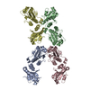 4tw5C  2b5eS  3t59S C: citing same article ( S: Starting model for refinement |
|---|---|
| Similar structure data |

-Links
 PDBj
PDBj

- Assembly
| Deposited unit | 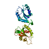
| ||||||||
|---|---|---|---|---|---|---|---|---|---|
| 1 |
| ||||||||
| Unit cell |
|
-Components
| #1: Protein | Mass: 31554.703 Da / Num. of mol.: 1 / Fragment: UNP residues 32-294 Source method: isolated from a genetically manipulated source Source: (gene. exp.) Naumovozyma dairenensis (fungus)Strain: ATCC 10597 / BCRC 20456 / CBS 421 / NBRC 0211 / NRRL Y-12639 Gene: NDAI0B01070, NDAI_0B01070 / Plasmid: pET15b / Production host:  Escherichia coli (E. coli) / Strain (production host): Origami B / References: UniProt: G0W5T0, EC: 5.4.3.1 Escherichia coli (E. coli) / Strain (production host): Origami B / References: UniProt: G0W5T0, EC: 5.4.3.1 |
|---|---|
| #2: Water | ChemComp-HOH / Water Mass: 18.015 Da / Num. of mol.: 351 / Source method: isolated from a natural source / Formula: H2O Mass: 18.015 Da / Num. of mol.: 351 / Source method: isolated from a natural source / Formula: H2O |
-Experimental details
-Experiment
| Experiment | Method: X-RAY DIFFRACTION / Number of used crystals: 1 |
|---|
- Sample preparation
| Crystal | Density Matthews: 2.56 Å3/Da / Density % sol: 52.01 % |
|---|---|
| Crystal grow | Temperature: 293 K / Method: vapor diffusion, sitting drop / pH: 6 Details: 25% w/v PEG 8,000, 0.1 M MES buffer, pH 6.0, 0.2 M calcium acetate |
-Data collection
| Diffraction | Mean temperature: 100 K |
|---|---|
| Diffraction source | Source: SYNCHROTRON / Site: ESRF  / Beamline: ID23-1 / Wavelength: 0.9763 Å / Beamline: ID23-1 / Wavelength: 0.9763 Å |
| Detector | Type: DECTRIS PILATUS 6M / Detector: PIXEL / Date: Dec 14, 2013 |
| Radiation | Protocol: SINGLE WAVELENGTH / Monochromatic (M) / Laue (L): M / Scattering type: x-ray |
| Radiation wavelength | Wavelength: 0.9763 Å / Relative weight: 1 |
| Reflection | Resolution: 1.8→50 Å / Num. obs: 30298 / % possible obs: 97.79 % / Redundancy: 8.2 % / Net I/σ(I): 9.8 |
| Reflection shell | Resolution: 1.8→1.86 Å / Redundancy: 8.1 % / Mean I/σ(I) obs: 2.9 / % possible all: 92.8 |
- Processing
| Software |
| ||||||||||||||||||||||||||||||||||||||||||||||||||||||||||||||||||||||||||||||||||||||||||||||||||||||||||||||||
|---|---|---|---|---|---|---|---|---|---|---|---|---|---|---|---|---|---|---|---|---|---|---|---|---|---|---|---|---|---|---|---|---|---|---|---|---|---|---|---|---|---|---|---|---|---|---|---|---|---|---|---|---|---|---|---|---|---|---|---|---|---|---|---|---|---|---|---|---|---|---|---|---|---|---|---|---|---|---|---|---|---|---|---|---|---|---|---|---|---|---|---|---|---|---|---|---|---|---|---|---|---|---|---|---|---|---|---|---|---|---|---|---|---|
| Refinement | Method to determine structure: MOLECULAR REPLACEMENT Starting model: 2B5E, 3T59 Resolution: 1.8→40.181 Å / SU ML: 0.18 / Cross valid method: FREE R-VALUE / σ(F): 1.36 / Phase error: 21.65 / Stereochemistry target values: ML
| ||||||||||||||||||||||||||||||||||||||||||||||||||||||||||||||||||||||||||||||||||||||||||||||||||||||||||||||||
| Solvent computation | Shrinkage radii: 0.9 Å / VDW probe radii: 1.11 Å / Solvent model: FLAT BULK SOLVENT MODEL | ||||||||||||||||||||||||||||||||||||||||||||||||||||||||||||||||||||||||||||||||||||||||||||||||||||||||||||||||
| Refinement step | Cycle: LAST / Resolution: 1.8→40.181 Å
| ||||||||||||||||||||||||||||||||||||||||||||||||||||||||||||||||||||||||||||||||||||||||||||||||||||||||||||||||
| Refine LS restraints |
| ||||||||||||||||||||||||||||||||||||||||||||||||||||||||||||||||||||||||||||||||||||||||||||||||||||||||||||||||
| LS refinement shell |
|
