 Movie
Movie Controller


+ Open data

- Basic information
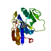
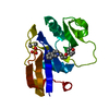
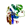
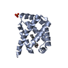
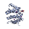
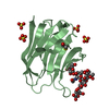
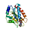
| Entry | Database: PDB / ID: 1uuh | ||||||
|---|---|---|---|---|---|---|---|
| Title | Hyaluronan binding domain of human CD44 | ||||||
 Components Components | CD44 ANTIGEN | ||||||
 Keywords Keywords |  LECTIN / HYALURONAN / EXTRACELLULAR MATRIX / RECEPTOR / LINK-DOMAIN / C- TYPE LECTIN / SUGAR-BINDING LECTIN / HYALURONAN / EXTRACELLULAR MATRIX / RECEPTOR / LINK-DOMAIN / C- TYPE LECTIN / SUGAR-BINDING | ||||||
| Function / homology |  Function and homology information Function and homology informationHyaluronan uptake and degradation / positive regulation of monocyte aggregation / hyaluronic acid binding / macrophage migration inhibitory factor receptor complex / regulation of lamellipodium morphogenesis / hyaluronan catabolic process / monocyte aggregation / cellular response to fibroblast growth factor stimulus / cartilage development / positive regulation of heterotypic cell-cell adhesion ...Hyaluronan uptake and degradation / positive regulation of monocyte aggregation / hyaluronic acid binding / macrophage migration inhibitory factor receptor complex / regulation of lamellipodium morphogenesis / hyaluronan catabolic process / monocyte aggregation / cellular response to fibroblast growth factor stimulus / cartilage development / positive regulation of heterotypic cell-cell adhesion / wound healing, spreading of cells / negative regulation of intrinsic apoptotic signaling pathway in response to DNA damage by p53 class mediator / negative regulation of DNA damage response, signal transduction by p53 class mediator / microvillus / lamellipodium membrane / Integrin cell surface interactions / collagen binding / Degradation of the extracellular matrix / T cell activation / cell-matrix adhesion / secretory granule membrane / cell projection / Cell surface interactions at the vascular wall / negative regulation of cysteine-type endopeptidase activity involved in apoptotic process / cell-cell adhesion / cytokine-mediated signaling pathway / Interferon gamma signaling / positive regulation of peptidyl-tyrosine phosphorylation / cell migration / transmembrane signaling receptor activity / positive regulation of peptidyl-serine phosphorylation / basolateral plasma membrane / positive regulation of ERK1 and ERK2 cascade / cell adhesion / inflammatory response / apical plasma membrane / focal adhesion / Neutrophil degranulation / negative regulation of apoptotic process / Golgi apparatus / cell surface / extracellular exosome / plasma membrane / cytosolSimilarity search - Function | ||||||
| Biological species |  HOMO SAPIENS (human) HOMO SAPIENS (human) | ||||||
| Method | X-RAY DIFFRACTION / SYNCHROTRON / OTHER / Resolution: 2.2 Å | ||||||
 Authors Authors | Teriete, P. / Banerji, S. / Noble, M. / Blundell, C. / Wright, A. / Pickford, A. / Lowe, E. / Mahoney, D. / Tammi, M. / Kahmann, J. ...Teriete, P. / Banerji, S. / Noble, M. / Blundell, C. / Wright, A. / Pickford, A. / Lowe, E. / Mahoney, D. / Tammi, M. / Kahmann, J. / Campbell, I. / Day, A. / Jackson, D. | ||||||
 Citation Citation | Journal: Mol.Cell / Year: 2004 Title: Structure of the Regulatory Hyaluronan-Binding Domain in the Inflammatory Leukocyte Homing Receptor Cd44 Authors: Teriete, P. / Banerji, S. / Noble, M. / Blundell, C. / Wright, A. / Pickford, A. / Lowe, E. / Mahoney, D. / Tammi, M. / Kahmann, J. / Campbell, I. / Day, A. / Jackson, D. | ||||||
| History |
|
- Structure visualization
| Structure viewer | Molecule: MolmilJmol/JSmol |
|---|
- Downloads & links
-Download
| PDBx/mmCIF format | 1uuh.cif.gz | 72.6 KB | Display | PDBx/mmCIF format |
|---|---|---|---|---|
| PDB format | pdb1uuh.ent.gz | 57.5 KB | Display | PDB format |
| PDBx/mmJSON format | 1uuh.json.gz | Tree view | PDBx/mmJSON format | |
| Others |  Other downloads Other downloads |
-Validation report
| Arichive directory | https://data.pdbj.org/pub/pdb/validation_reports/uu/1uuhftp://data.pdbj.org/pub/pdb/validation_reports/uu/1uuh | HTTPS FTP |
|---|
-Related structure data


-Links
 PDBj
PDBj


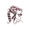
- Assembly
| Deposited unit | 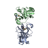
| ||||||||
|---|---|---|---|---|---|---|---|---|---|
| 1 | 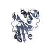
| ||||||||
| 2 | 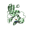
| ||||||||
| Unit cell |
| ||||||||
| Noncrystallographic symmetry (NCS) | NCS oper: (Code: given Matrix: (-0.74746, -0.61772, -0.24439), Vector : |
-Components
| #1: Protein | Mass: 17695.275 Da / Num. of mol.: 2 / Fragment: HYALURONAN BINDING DOMAIN, RESIDUES 20-178 Source method: isolated from a genetically manipulated source Details: FIRST RESIDUE IS IN FACT THE LAST RESIDUE OF THE SIGNAL PEPTIDE, AND WOULD NORMALLY BE CLEAVED Source: (gene. exp.) HOMO SAPIENS (human) / Plasmid: PET19B / Production host:  ESCHERICHIA COLI (E. coli) / Strain (production host): BL21 / Variant (production host): DE3 PLYSS / References: UniProt: P16070 ESCHERICHIA COLI (E. coli) / Strain (production host): BL21 / Variant (production host): DE3 PLYSS / References: UniProt: P16070#2: Water | ChemComp-HOH / | Water Mass: 18.015 Da / Num. of mol.: 262 / Source method: isolated from a natural source / Formula: H2O Mass: 18.015 Da / Num. of mol.: 262 / Source method: isolated from a natural source / Formula: H2OSequence details | HYALURONAN | |
|---|
-Experimental details
-Experiment
| Experiment | Method: X-RAY DIFFRACTION / Number of used crystals: 1 |
|---|
- Sample preparation
| Crystal | Density Matthews: 2.3 Å3/Da / Density % sol: 40 % | ||||||||||||||||||||||||
|---|---|---|---|---|---|---|---|---|---|---|---|---|---|---|---|---|---|---|---|---|---|---|---|---|---|
| Crystal grow | Method: vapor diffusion, hanging drop / pH: 5.5 Details: HANGING-DROP PROTEIN 13 MG/ML; WELL BUFFER 12% PEG 3350, 50MM NACL, 10% GLYCEROL, pH 5.50 | ||||||||||||||||||||||||
| Crystal grow | *PLUS Method: vapor diffusion, hanging drop | ||||||||||||||||||||||||
| Components of the solutions | *PLUS
|
-Data collection
| Diffraction | Mean temperature: 100 K |
|---|---|
| Diffraction source | Source: SYNCHROTRON / Site: ESRF  / Beamline: ID14-4 / Wavelength: 0.9795 / Beamline: ID14-4 / Wavelength: 0.9795 |
| Detector | Type: ADSC CCD / Detector: CCD / Date: May 15, 2002 |
| Radiation | Protocol: SINGLE WAVELENGTH / Monochromatic (M) / Laue (L): M / Scattering type: x-ray |
| Radiation wavelength | Wavelength: 0.9795 Å / Relative weight: 1 |
| Reflection | Resolution: 2.2→23.31 Å / Num. obs: 17476 / % possible obs: 99.8 % / Redundancy: 3.82 % / Rmerge(I) obs: 0.076 / Net I/σ(I): 7.2989 |
| Reflection shell | Resolution: 2.2→2.32 Å / Redundancy: 3.53 % / Rmerge(I) obs: 0.18 / Mean I/σ(I) obs: 3.16 / % possible all: 99.6 |
| Reflection | *PLUS Highest resolution: 2.2 Å / Lowest resolution: 23.31 Å / % possible obs: 99.4 % / Redundancy: 3.8 % / Rmerge(I) obs: 0.076 |
| Reflection shell | *PLUS % possible obs: 98.2 % / Redundancy: 3.5 % / Rmerge(I) obs: 0.181 / Mean I/σ(I) obs: 3.1 |
- Processing
| Software |
| ||||||||||||||||||||||||||||||||||||||||||||||||||||||||||||||||||||||||||||||||||||||||||||||||||||||||||||||||||||||||||||||||||||||||||||||||||||||||||||||||||||||||||||||||||||||
|---|---|---|---|---|---|---|---|---|---|---|---|---|---|---|---|---|---|---|---|---|---|---|---|---|---|---|---|---|---|---|---|---|---|---|---|---|---|---|---|---|---|---|---|---|---|---|---|---|---|---|---|---|---|---|---|---|---|---|---|---|---|---|---|---|---|---|---|---|---|---|---|---|---|---|---|---|---|---|---|---|---|---|---|---|---|---|---|---|---|---|---|---|---|---|---|---|---|---|---|---|---|---|---|---|---|---|---|---|---|---|---|---|---|---|---|---|---|---|---|---|---|---|---|---|---|---|---|---|---|---|---|---|---|---|---|---|---|---|---|---|---|---|---|---|---|---|---|---|---|---|---|---|---|---|---|---|---|---|---|---|---|---|---|---|---|---|---|---|---|---|---|---|---|---|---|---|---|---|---|---|---|---|---|
| Refinement | Method to determine structure: OTHER / Resolution: 2.2→57.73 Å / Cor.coef. Fo:Fc: 0.933 / Cor.coef. Fo:Fc free: 0.878 / SU B: 5.483 / SU ML: 0.141 / Cross valid method: THROUGHOUT / ESU R: 0.261 / ESU R Free: 0.216 / Stereochemistry target values: MAXIMUM LIKELIHOOD
| ||||||||||||||||||||||||||||||||||||||||||||||||||||||||||||||||||||||||||||||||||||||||||||||||||||||||||||||||||||||||||||||||||||||||||||||||||||||||||||||||||||||||||||||||||||||
| Solvent computation | Ion probe radii: 0.8 Å / Shrinkage radii: 0.8 Å / VDW probe radii: 1.4 Å / Solvent model: BABINET MODEL PLUS MASK | ||||||||||||||||||||||||||||||||||||||||||||||||||||||||||||||||||||||||||||||||||||||||||||||||||||||||||||||||||||||||||||||||||||||||||||||||||||||||||||||||||||||||||||||||||||||
| Displacement parameters | Biso mean: 17.45 Å2
| ||||||||||||||||||||||||||||||||||||||||||||||||||||||||||||||||||||||||||||||||||||||||||||||||||||||||||||||||||||||||||||||||||||||||||||||||||||||||||||||||||||||||||||||||||||||
| Refinement step | Cycle: LAST / Resolution: 2.2→57.73 Å
| ||||||||||||||||||||||||||||||||||||||||||||||||||||||||||||||||||||||||||||||||||||||||||||||||||||||||||||||||||||||||||||||||||||||||||||||||||||||||||||||||||||||||||||||||||||||
| Refine LS restraints |
|