- PDB-3l81: Crystal structure of adaptor protein complex 4 (AP-4) mu4 subunit... -
+
Open data
ID or keywords:
Loading...
-
Basic information
Entry
Database: PDB / ID: 3l81
Title
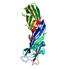
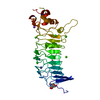
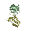
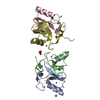
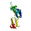
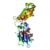
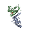
Crystal structure of adaptor protein complex 4 (AP-4) mu4 subunit C-terminal domain, in complex with a sorting peptide from the amyloid precursor protein (APP)
Components
AP-4 complex subunit mu-1
Amyloid beta A4 protein
Keywords
TRANSPORT PROTEIN / immunoglobulin-like beta-sandwich / Coated pit / Golgi apparatus / Membrane / Phosphoprotein / Protein transport / Transport / Alzheimer disease / Amyloid / Amyloidosis
Function / homology
Function and homology information
AP-4 adaptor complex / protein localization to basolateral plasma membrane / Golgi to lysosome transport / clathrin adaptor complex / post-Golgi vesicle-mediated transport / Golgi to endosome transport / protein targeting to lysosome / regulation of epidermal growth factor-activated receptor activity / signaling receptor activator activity / cytosolic mRNA polyadenylation ...AP-4 adaptor complex / protein localization to basolateral plasma membrane / Golgi to lysosome transport / clathrin adaptor complex / post-Golgi vesicle-mediated transport / Golgi to endosome transport / protein targeting to lysosome / regulation of epidermal growth factor-activated receptor activity / signaling receptor activator activity / cytosolic mRNA polyadenylation / collateral sprouting in absence of injury / microglia development / regulation of synapse structure or activity / Formyl peptide receptors bind formyl peptides and many other ligands / regulation of Wnt signaling pathway / axo-dendritic transport / synaptic assembly at neuromuscular junction / smooth endoplasmic reticulum calcium ion homeostasis / axon midline choice point recognition / astrocyte activation involved in immune response / regulation of spontaneous synaptic transmission / mating behavior / NMDA selective glutamate receptor signaling pathway / ciliary rootlet / Lysosome Vesicle Biogenesis / PTB domain binding / Golgi-associated vesicle / positive regulation of amyloid fibril formation / neuron remodeling / Insertion of tail-anchored proteins into the endoplasmic reticulum membrane / protein serine/threonine kinase binding / Deregulated CDK5 triggers multiple neurodegenerative pathways in Alzheimer's disease models / : / nuclear envelope lumen / suckling behavior / presynaptic active zone / dendrite development / COPII-coated ER to Golgi transport vesicle / modulation of excitatory postsynaptic potential / TRAF6 mediated NF-kB activation / Advanced glycosylation endproduct receptor signaling / neuromuscular process controlling balance / The NLRP3 inflammasome / autophagosome assembly / regulation of presynapse assembly / transition metal ion binding / protein transmembrane transporter activity / regulation of multicellular organism growth / intracellular copper ion homeostasis / negative regulation of long-term synaptic potentiation / negative regulation of neuron differentiation / ECM proteoglycans / smooth endoplasmic reticulum / Mitochondrial protein degradation / positive regulation of T cell migration / spindle midzone / protein targeting / Purinergic signaling in leishmaniasis infection / positive regulation of calcium-mediated signaling / clathrin-coated pit / regulation of peptidyl-tyrosine phosphorylation / positive regulation of chemokine production / vesicle-mediated transport / forebrain development / Notch signaling pathway / positive regulation of G2/M transition of mitotic cell cycle / neuron projection maintenance / positive regulation of protein metabolic process / ionotropic glutamate receptor signaling pathway / positive regulation of glycolytic process / cholesterol metabolic process / response to interleukin-1 / positive regulation of mitotic cell cycle / extracellular matrix organization / axonogenesis / adult locomotory behavior / trans-Golgi network membrane / platelet alpha granule lumen / locomotory behavior / positive regulation of peptidyl-threonine phosphorylation / dendritic shaft / learning / positive regulation of interleukin-1 beta production / central nervous system development / positive regulation of long-term synaptic potentiation / endosome lumen / astrocyte activation / Post-translational protein phosphorylation / positive regulation of JNK cascade / synapse organization / regulation of long-term neuronal synaptic plasticity / intracellular protein transport / microglial cell activation / TAK1-dependent IKK and NF-kappa-B activation / visual learning / trans-Golgi network / serine-type endopeptidase inhibitor activity / neuromuscular junction / protein localization / recycling endosome Similarity search - Function
Mu homology domain, subdomain B / Clathrin adaptor, mu subunit / Clathrin adaptor, mu subunit, conserved site / Clathrin adaptor complexes medium chain signature 2. / Adaptor complexes medium subunit family / AP complex, mu/sigma subunit / Clathrin adaptor complex small chain / AP-2 complex subunit mu, C-terminal superfamily / Mu homology domain / Mu homology domain (MHD) profile. ...Mu homology domain, subdomain B / Clathrin adaptor, mu subunit / Clathrin adaptor, mu subunit, conserved site / Clathrin adaptor complexes medium chain signature 2. / Adaptor complexes medium subunit family / AP complex, mu/sigma subunit / Clathrin adaptor complex small chain / AP-2 complex subunit mu, C-terminal superfamily / Mu homology domain / Mu homology domain (MHD) profile. / Longin-like domain superfamily / Amyloidogenic glycoprotein, copper-binding / Amyloidogenic glycoprotein, copper-binding domain conserved site / Amyloidogenic glycoprotein, copper-binding domain superfamily / Copper-binding of amyloid precursor, CuBD / Amyloid precursor protein (APP) copper-binding (CuBD) domain signature. / Amyloidogenic glycoprotein, amyloid-beta peptide superfamily / Beta-amyloid peptide (beta-APP) / Amyloidogenic glycoprotein, amyloid-beta peptide / Beta-amyloid precursor protein C-terminal / Amyloidogenic glycoprotein, intracellular domain, conserved site / Beta-amyloid precursor protein C-terminus / Amyloid precursor protein (APP) intracellular domain signature. / Amyloid precursor protein (APP) E1 domain profile. / Amyloid precursor protein (APP) E2 domain profile. / Amyloidogenic glycoprotein, extracellular / Amyloidogenic glycoprotein, heparin-binding / Amyloidogenic glycoprotein, E2 domain / E2 domain superfamily / Amyloidogenic glycoprotein, heparin-binding domain superfamily / Amyloid A4 N-terminal heparin-binding / E2 domain of amyloid precursor protein / amyloid A4 / Amyloidogenic glycoprotein / Proteinase inhibitor I2, Kunitz, conserved site / Pancreatic trypsin inhibitor (Kunitz) family signature. / BPTI/Kunitz family of serine protease inhibitors. / Pancreatic trypsin inhibitor Kunitz domain / Kunitz/Bovine pancreatic trypsin inhibitor domain / Pancreatic trypsin inhibitor (Kunitz) family profile. / Pancreatic trypsin inhibitor Kunitz domain superfamily / PH-like domain superfamily / Immunoglobulin-like / Sandwich / Mainly Beta Similarity search - Domain/homology
In the structure databanks used in Yorodumi, some data are registered as the other names, "COVID-19 virus" and "2019-nCoV". Here are the details of the virus and the list of structure data.
Jan 31, 2019. EMDB accession codes are about to change! (news from PDBe EMDB page)
EMDB accession codes are about to change! (news from PDBe EMDB page)
The allocation of 4 digits for EMDB accession codes will soon come to an end. Whilst these codes will remain in use, new EMDB accession codes will include an additional digit and will expand incrementally as the available range of codes is exhausted. The current 4-digit format prefixed with “EMD-” (i.e. EMD-XXXX) will advance to a 5-digit format (i.e. EMD-XXXXX), and so on. It is currently estimated that the 4-digit codes will be depleted around Spring 2019, at which point the 5-digit format will come into force.
The EM Navigator/Yorodumi systems omit the EMD- prefix.
Related info.:Q: What is EMD? / ID/Accession-code notation in Yorodumi/EM Navigator
Yorodumi is a browser for structure data from EMDB, PDB, SASBDB, etc.
This page is also the successor to EM Navigator detail page, and also detail information page/front-end page for Omokage search.
The word "yorodu" (or yorozu) is an old Japanese word meaning "ten thousand". "mi" (miru) is to see.
Related info.:EMDB / PDB / SASBDB / Comparison of 3 databanks / Yorodumi Search / Aug 31, 2016. New EM Navigator & Yorodumi / Yorodumi Papers / Jmol/JSmol / Function and homology information / Changes in new EM Navigator and Yorodumi
 Movie
Movie Controller

 Yorodumi
 Open data
 Basic information
 Components
 Keywords
 TRANSPORT PROTEIN / immunoglobulin-like beta-sandwich / Coated pit /
  Function and homology information
 Authors
 Citation
 Structure visualization
 Downloads & links
 Other downloads


 PDBj
PDBj


 Assembly
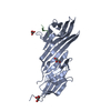


 Mass: 92.094 Da / Num. of mol.: 3 / Source method: obtained synthetically / Formula: C3H8O3
 Mass: 18.015 Da / Num. of mol.: 153 / Source method: isolated from a natural source / Formula: H2O
 Sample preparation
 Processing