 Movie
Movie Controller


+ Open data

- Basic information
| Entry | Database: PDB / ID: 6sqp | |||||||||
|---|---|---|---|---|---|---|---|---|---|---|
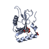
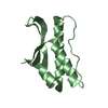
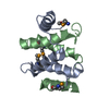
| Title | Crystal structure of Cat MDM2-S429E RING domain homodimer | |||||||||
 Components Components | (E3 ubiquitin-protein ligase ...) x 3 | |||||||||
 Keywords Keywords |  LIGASE / MDM2 / MDMX / ubiquitin ligase / E3 / phosphorylation LIGASE / MDM2 / MDMX / ubiquitin ligase / E3 / phosphorylation | |||||||||
| Function / homology |  Function and homology information Function and homology informationregulation of biological quality / ribonucleoprotein complex binding / ubiquitin binding / RING-type E3 ubiquitin transferase / ubiquitin protein ligase activity / p53 binding / 5S rRNA binding / regulation of gene expression / protein ubiquitination / regulation of cell cycle ...regulation of biological quality / ribonucleoprotein complex binding / ubiquitin binding / RING-type E3 ubiquitin transferase / ubiquitin protein ligase activity / p53 binding / 5S rRNA binding / regulation of gene expression / protein ubiquitination / regulation of cell cycle / cell cycle / negative regulation of DNA-templated transcription / apoptotic process / nucleolus / negative regulation of apoptotic process / negative regulation of transcription by RNA polymerase II / nucleoplasm / identical protein binding / metal ion binding / nucleus / cytoplasmSimilarity search - Function | |||||||||
| Biological species |  Felis catus (domestic cat) Felis catus (domestic cat) | |||||||||
| Method | X-RAY DIFFRACTION / SYNCHROTRON / MOLECULAR REPLACEMENT / Resolution: 1.21 Å | |||||||||
 Authors Authors | Magnussen, H.M. / Ahmed, S.F. / Huang, D.T. | |||||||||
| Funding support |  United Kingdom, United Kingdom,  Belgium, 2items Belgium, 2items
| |||||||||
 Citation Citation | Journal: Nat Commun / Year: 2020 Title: Structural basis for DNA damage-induced phosphoregulation of MDM2 RING domain. Authors: Magnussen, H.M. / Ahmed, S.F. / Sibbet, G.J. / Hristova, V.A. / Nomura, K. / Hock, A.K. / Archibald, L.J. / Jamieson, A.G. / Fushman, D. / Vousden, K.H. / Weissman, A.M. / Huang, D.T. | |||||||||
| History |
|
- Structure visualization
| Structure viewer | Molecule: MolmilJmol/JSmol |
|---|
- Downloads & links
-Download
| PDBx/mmCIF format | 6sqp.cif.gz | 127.2 KB | Display | PDBx/mmCIF format |
|---|---|---|---|---|
| PDB format | pdb6sqp.ent.gz | 97.9 KB | Display | PDB format |
| PDBx/mmJSON format | 6sqp.json.gz | Tree view | PDBx/mmJSON format | |
| Others |  Other downloads Other downloads |
-Validation report
| Arichive directory | https://data.pdbj.org/pub/pdb/validation_reports/sq/6sqpftp://data.pdbj.org/pub/pdb/validation_reports/sq/6sqp | HTTPS FTP |
|---|
-Related structure data
| Related structure data |  6sqoC  6sqrC  6sqsC  5mnjS S: Starting model for refinement C: citing same article ( |
|---|---|
| Similar structure data |


-Links
 PDBj
PDBj

- Assembly
| Deposited unit | 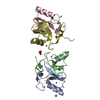
| ||||||||
|---|---|---|---|---|---|---|---|---|---|
| 1 | 
| ||||||||
| 2 | 
| ||||||||
| Unit cell |
|
-Components
-E3 ubiquitin-protein ligase ... , 3 types, 4 molecules ABDC
| #1: Protein | Mass: 7152.696 Da / Num. of mol.: 1 / Mutation: S429E, G443T Source method: isolated from a genetically manipulated source Details: Residues 422-491 and contains S429E and G443T mutation. GS at the N-terminus resulted from cloning. Source: (gene. exp.) Felis catus (domestic cat) / Gene: MDM2 / Production host:  Escherichia coli (E. coli) Escherichia coli (E. coli)References: UniProt: Q7YRZ8, RING-type E3 ubiquitin transferase | ||
|---|---|---|---|
| #2: Protein | Mass: 7720.328 Da / Num. of mol.: 2 / Mutation: S429E, G443T Source method: isolated from a genetically manipulated source Details: Residues 422-491 and contains S429E and G443T. GS at the N-terminus resulted from cloning. Source: (gene. exp.) Felis catus (domestic cat) / Gene: MDM2 / Production host: Escherichia coli (E. coli)References: UniProt: Q7YRZ8, RING-type E3 ubiquitin transferase #3: Protein | | Mass: 7378.926 Da / Num. of mol.: 1 / Mutation: S429E, G443T Source method: isolated from a genetically manipulated source Details: Residues 422-491 and contains S429E and G443T. GS at the N-terminus resulted from cloning. Source: (gene. exp.) Felis catus (domestic cat) / Gene: MDM2 / Production host: Escherichia coli (E. coli)References: UniProt: Q7YRZ8, RING-type E3 ubiquitin transferase |
-Non-polymers , 4 types, 254 molecules 


| #4: Chemical | ChemComp-ZN /  Mass: 65.409 Da / Num. of mol.: 8 / Source method: obtained synthetically / Formula: Zn Mass: 65.409 Da / Num. of mol.: 8 / Source method: obtained synthetically / Formula: Zn#5: Chemical | Chloride Mass: 35.453 Da / Num. of mol.: 2 / Source method: obtained synthetically / Formula: Cl Mass: 35.453 Da / Num. of mol.: 2 / Source method: obtained synthetically / Formula: Cl#6: Chemical | ChemComp-NO3 / | Nitrate Mass: 62.005 Da / Num. of mol.: 1 / Source method: obtained synthetically / Formula: NO3 Mass: 62.005 Da / Num. of mol.: 1 / Source method: obtained synthetically / Formula: NO3#7: Water | ChemComp-HOH / | WaterMass: 18.015 Da / Num. of mol.: 243 / Source method: isolated from a natural source / Formula: H2O |
|---|
-Details
| Has ligand of interest | N |
|---|
-Experimental details
-Experiment
| Experiment | Method: X-RAY DIFFRACTION / Number of used crystals: 1 |
|---|
- Sample preparation
| Crystal | Density Matthews: 2.02 Å3/Da / Density % sol: 39.13 % |
|---|---|
| Crystal grow | Temperature: 292 K / Method: vapor diffusion / Details: 0.1 M MMT, pH 9.0 and 25% (w/v) PEG1500 |
-Data collection
| Diffraction | Mean temperature: 100 K / Serial crystal experiment: N |
|---|---|
| Diffraction source | Source: SYNCHROTRON / Site: Diamond / Beamline: I04-1 / Wavelength: 0.916 Å |
| Detector | Type: DECTRIS PILATUS 6M-F / Detector: PIXEL / Date: Jul 7, 2018 |
| Radiation | Protocol: SINGLE WAVELENGTH / Monochromatic (M) / Laue (L): M / Scattering type: x-ray |
| Radiation wavelength | Wavelength: 0.916 Å / Relative weight: 1 |
| Reflection | Resolution: 1.21→23.53 Å / Num. obs: 71796 / % possible obs: 98.3 % / Redundancy: 3.2 % / CC1/2: 0.991 / Net I/σ(I): 5.1 |
| Reflection shell | Resolution: 1.21→1.24 Å / Num. unique obs: 5274 / CC1/2: 0.608 |
- Processing
| Software |
| ||||||||||||||||
|---|---|---|---|---|---|---|---|---|---|---|---|---|---|---|---|---|---|
| Refinement | Method to determine structure: MOLECULAR REPLACEMENT Starting model: 5MNJ Resolution: 1.21→23.53 Å / Cross valid method: FREE R-VALUE
| ||||||||||||||||
| Refinement step | Cycle: LAST / Resolution: 1.21→23.53 Å
|
