 Movie
Movie Controller

[English] 日本語
 Yorodumi
- PDB-6sqo: Crystal structure of human MDM2 RING domain homodimer bound to Ub... -
+ Open data

- Basic information
| Entry | Database: PDB / ID: 6sqo | |||||||||
|---|---|---|---|---|---|---|---|---|---|---|
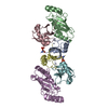
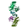
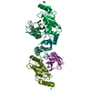
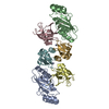
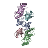
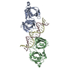
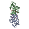
| Title | Crystal structure of human MDM2 RING domain homodimer bound to UbcH5B-Ub | |||||||||
 Components Components |
| |||||||||
 Keywords Keywords |  LIGASE / Ubiquitin ligase / E3 / MDM2 / MDMX / E2 / phosphorylation / p53 LIGASE / Ubiquitin ligase / E3 / MDM2 / MDMX / E2 / phosphorylation / p53 | |||||||||
| Function / homology |  Function and homology information Function and homology informationcellular response to vitamin B1 / response to formaldehyde / response to water-immersion restraint stress / (E3-independent) E2 ubiquitin-conjugating enzyme / traversing start control point of mitotic cell cycle / negative regulation of intrinsic apoptotic signaling pathway by p53 class mediator / response to ether / negative regulation of signal transduction by p53 class mediator / fibroblast activation / atrial septum development ...cellular response to vitamin B1 / response to formaldehyde / response to water-immersion restraint stress / (E3-independent) E2 ubiquitin-conjugating enzyme / traversing start control point of mitotic cell cycle / negative regulation of intrinsic apoptotic signaling pathway by p53 class mediator / response to ether / negative regulation of signal transduction by p53 class mediator / fibroblast activation / atrial septum development / receptor serine/threonine kinase binding / Trafficking of AMPA receptors / positive regulation of vascular associated smooth muscle cell migration / peroxisome proliferator activated receptor binding / response to iron ion / negative regulation of protein processing / Formation of the ternary complex, and subsequently, the 43S complex / SUMO transferase activity / response to steroid hormone / NEDD8 ligase activity / AKT phosphorylates targets in the cytosol / atrioventricular valve morphogenesis / cellular response to peptide hormone stimulus / Ribosomal scanning and start codon recognition / ventricular septum development / endocardial cushion morphogenesis / Translation initiation complex formation / E2 ubiquitin-conjugating enzyme / positive regulation of muscle cell differentiation / SUMOylation of ubiquitinylation proteins / cellular response to alkaloid / blood vessel development / regulation of protein catabolic process / cardiac septum morphogenesis / SARS-CoV-1 modulates host translation machinery / Constitutive Signaling by AKT1 E17K in Cancer / Peptide chain elongation / Selenocysteine synthesis / ligase activity / Formation of a pool of free 40S subunits / negative regulation of DNA damage response, signal transduction by p53 class mediator / ubiquitin conjugating enzyme activity / Eukaryotic Translation Termination / response to magnesium ion / Response of EIF2AK4 (GCN2) to amino acid deficiency / protein sumoylation / SUMOylation of transcription factors / SRP-dependent cotranslational protein targeting to membrane / Viral mRNA Translation / protein localization to nucleus / Nonsense Mediated Decay (NMD) independent of the Exon Junction Complex (EJC) / GTP hydrolysis and joining of the 60S ribosomal subunit / L13a-mediated translational silencing of Ceruloplasmin expression / cellular response to UV-C / Major pathway of rRNA processing in the nucleolus and cytosol / blood vessel remodeling / cellular response to estrogen stimulus / protein autoubiquitination / Nonsense Mediated Decay (NMD) enhanced by the Exon Junction Complex (EJC) / protein K48-linked ubiquitination / cellular response to actinomycin D / ribonucleoprotein complex binding / cytosolic ribosome / ubiquitin ligase complex / Maturation of protein E / Maturation of protein E / ER Quality Control Compartment (ERQC) / Myoclonic epilepsy of Lafora / FLT3 signaling by CBL mutants / Prevention of phagosomal-lysosomal fusion / IRAK2 mediated activation of TAK1 complex / Alpha-protein kinase 1 signaling pathway / Glycogen synthesis / positive regulation of vascular associated smooth muscle cell proliferation / IRAK1 recruits IKK complex / IRAK1 recruits IKK complex upon TLR7/8 or 9 stimulation / Membrane binding and targetting of GAG proteins / Endosomal Sorting Complex Required For Transport (ESCRT) / IRAK2 mediated activation of TAK1 complex upon TLR7/8 or 9 stimulation / PTK6 Regulates RTKs and Their Effectors AKT1 and DOK1 / Negative regulation of FLT3 / Constitutive Signaling by NOTCH1 HD Domain Mutants / Regulation of FZD by ubiquitination / TICAM1,TRAF6-dependent induction of TAK1 complex / NOTCH2 Activation and Transmission of Signal to the Nucleus / TICAM1-dependent activation of IRF3/IRF7 / APC/C:Cdc20 mediated degradation of Cyclin B / DNA damage response, signal transduction by p53 class mediator resulting in cell cycle arrest / p75NTR recruits signalling complexes / Downregulation of ERBB4 signaling / TRAF6 mediated IRF7 activation in TLR7/8 or 9 signaling / APC-Cdc20 mediated degradation of Nek2A / PINK1-PRKN Mediated Mitophagy / NPAS4 regulates expression of target genes / TRAF6-mediated induction of TAK1 complex within TLR4 complex / Pexophagy / Regulation of innate immune responses to cytosolic DNA / transcription repressor complex / VLDLR internalisation and degradation / InlA-mediated entry of Listeria monocytogenes into host cellsSimilarity search - Function | |||||||||
| Biological species |  Homo sapiens (human) Homo sapiens (human) | |||||||||
| Method | X-RAY DIFFRACTION / SYNCHROTRON / MOLECULAR REPLACEMENT / Resolution: 1.41 Å | |||||||||
 Authors Authors | Magnussen, H.M. / Ahmed, S.F. / Huang, D.T. | |||||||||
| Funding support |  United Kingdom, United Kingdom,  Belgium, 2items Belgium, 2items
| |||||||||
 Citation Citation | Journal: Nat Commun / Year: 2020 Title: Structural basis for DNA damage-induced phosphoregulation of MDM2 RING domain. Authors: Magnussen, H.M. / Ahmed, S.F. / Sibbet, G.J. / Hristova, V.A. / Nomura, K. / Hock, A.K. / Archibald, L.J. / Jamieson, A.G. / Fushman, D. / Vousden, K.H. / Weissman, A.M. / Huang, D.T. | |||||||||
| History |
|
- Structure visualization
| Structure viewer | Molecule: MolmilJmol/JSmol |
|---|
- Downloads & links
-Download
| PDBx/mmCIF format | 6sqo.cif.gz | 266.9 KB | Display | PDBx/mmCIF format |
|---|---|---|---|---|
| PDB format | pdb6sqo.ent.gz | 214.9 KB | Display | PDB format |
| PDBx/mmJSON format | 6sqo.json.gz | Tree view | PDBx/mmJSON format | |
| Others |  Other downloads Other downloads |
-Validation report
| Arichive directory | https://data.pdbj.org/pub/pdb/validation_reports/sq/6sqoftp://data.pdbj.org/pub/pdb/validation_reports/sq/6sqo | HTTPS FTP |
|---|
-Related structure data
| Related structure data | 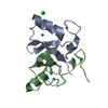 6sqpC  6sqrC  6sqsC  5mnjS S: Starting model for refinement C: citing same article ( |
|---|---|
| Similar structure data |


-Links
 PDBj
PDBj


- Assembly
| Deposited unit | 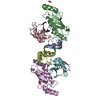
| ||||||||
|---|---|---|---|---|---|---|---|---|---|
| 1 |
| ||||||||
| Unit cell |
|
-Components
-Protein , 3 types, 6 molecules ADBECF
| #1: Protein | Mass: 6920.525 Da / Num. of mol.: 2 Source method: isolated from a genetically manipulated source Details: Construct contains MDM2 residues 419-491. Residues 419-427 are absent due to lack of electron density. GS at the N-terminus resulted from cloning. Source: (gene. exp.) Homo sapiens (human) / Gene: MDM2 / Production host:  Escherichia coli (E. coli) Escherichia coli (E. coli)References: UniProt: Q00987, RING-type E3 ubiquitin transferase #2: Protein | Mass: 16720.186 Da / Num. of mol.: 2 / Mutation: S22R, C85K Source method: isolated from a genetically manipulated source Details: K85 sidechains from Chains B and E form an isopeptide bond with the C-terminus of G76 in Chains C and F, respectively. Source: (gene. exp.) Homo sapiens (human) / Gene: UBE2D2, PUBC1, UBC4, UBC5B, UBCH4, UBCH5B / Production host: Escherichia coli (E. coli)References: UniProt: P62837, E2 ubiquitin-conjugating enzyme, (E3-independent) E2 ubiquitin-conjugating enzyme#3: Protein | Mass: 8663.908 Da / Num. of mol.: 2 Source method: isolated from a genetically manipulated source Details: GSGGS LINKER AT THE N-TERMINUS RESULTED FROM CLONING. G76 IN CHAINS C and F ARE COVALENTLY LINKED TO K85 SIDE CHAIN IN CHAINS B AND E, RESPECTIVELY. Source: (gene. exp.) Homo sapiens (human) / Gene: RPS27A / Production host: Escherichia coli (E. coli) / References: UniProt: J3QTR3, UniProt: P62979*PLUS |
|---|
-Non-polymers , 4 types, 556 molecules 


| #4: Chemical | ChemComp-ZN /  Mass: 65.409 Da / Num. of mol.: 4 / Source method: obtained synthetically / Formula: Zn Mass: 65.409 Da / Num. of mol.: 4 / Source method: obtained synthetically / Formula: Zn#5: Chemical | ChemComp-CL / Chloride Mass: 35.453 Da / Num. of mol.: 4 / Source method: obtained synthetically / Formula: Cl Mass: 35.453 Da / Num. of mol.: 4 / Source method: obtained synthetically / Formula: Cl#6: Chemical | Nitrate Mass: 62.005 Da / Num. of mol.: 2 / Source method: obtained synthetically / Formula: NO3 Mass: 62.005 Da / Num. of mol.: 2 / Source method: obtained synthetically / Formula: NO3#7: Water | ChemComp-HOH / | WaterMass: 18.015 Da / Num. of mol.: 546 / Source method: isolated from a natural source / Formula: H2O |
|---|
-Details
| Has ligand of interest | N |
|---|
-Experimental details
-Experiment
| Experiment | Method: X-RAY DIFFRACTION / Number of used crystals: 1 |
|---|
- Sample preparation
| Crystal | Density Matthews: 2.65 Å3/Da / Density % sol: 53.61 % |
|---|---|
| Crystal grow | Temperature: 292 K / Method: vapor diffusion Details: 0.1 M SPG, pH 7.0, 10% (w/v) PEG Smear Broad, 0.15 M NH4NO3 |
-Data collection
| Diffraction | Mean temperature: 100 K / Serial crystal experiment: N |
|---|---|
| Diffraction source | Source: SYNCHROTRON / Site: Diamond / Beamline: I04-1 / Wavelength: 0.916 Å |
| Detector | Type: DECTRIS PILATUS 6M-F / Detector: PIXEL / Date: May 6, 2018 |
| Radiation | Protocol: SINGLE WAVELENGTH / Monochromatic (M) / Laue (L): M / Scattering type: x-ray |
| Radiation wavelength | Wavelength: 0.916 Å / Relative weight: 1 |
| Reflection | Resolution: 1.41→112 Å / Num. obs: 129794 / % possible obs: 100 % / Redundancy: 8.9 % / CC1/2: 1 / Rmerge(I) obs: 0.047 / Net I/σ(I): 19.2 |
| Reflection shell | Resolution: 1.41→1.43 Å / Num. unique obs: 6423 / CC1/2: 0.542 |
- Processing
| Software |
| ||||||||||||||||
|---|---|---|---|---|---|---|---|---|---|---|---|---|---|---|---|---|---|
| Refinement | Method to determine structure: MOLECULAR REPLACEMENT Starting model: 5MNJ Resolution: 1.41→112 Å / Cross valid method: FREE R-VALUE
| ||||||||||||||||
| Refinement step | Cycle: LAST / Resolution: 1.41→112 Å
|
