 Movie
Movie Controller

[English] 日本語
 Yorodumi
- PDB-1bxx: MU2 ADAPTIN SUBUNIT (AP50) OF AP2 ADAPTOR (SECOND DOMAIN), COMPLE... -
+ Open data

- Basic information
| Entry | Database: PDB / ID: 1bxx | ||||||
|---|---|---|---|---|---|---|---|
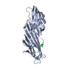
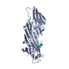
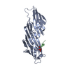
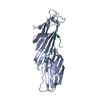
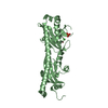
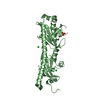
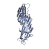
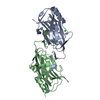
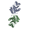
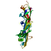
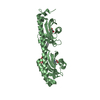
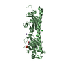
| Title | MU2 ADAPTIN SUBUNIT (AP50) OF AP2 ADAPTOR (SECOND DOMAIN), COMPLEXED WITH TGN38 INTERNALIZATION PEPTIDE DYQRLN | ||||||
 Components Components |
| ||||||
 Keywords Keywords | ENDOCYTOSIS/EXOCYTOSIS /  ENDOCYTOSIS / ADAPTOR / PEPTIDE COMPLEX / ENDOCYTOSIS-EXOCYTOSIS COMPLEX ENDOCYTOSIS / ADAPTOR / PEPTIDE COMPLEX / ENDOCYTOSIS-EXOCYTOSIS COMPLEX | ||||||
| Function / homology |  Function and homology information Function and homology informationGap junction degradation / Formation of annular gap junctions / WNT5A-dependent internalization of FZD2, FZD5 and ROR2 / LDL clearance / VLDLR internalisation and degradation / Retrograde neurotrophin signalling / WNT5A-dependent internalization of FZD4 / extrinsic component of presynaptic endocytic zone membrane / MHC class II antigen presentation / AP-2 adaptor complex ...Gap junction degradation / Formation of annular gap junctions / WNT5A-dependent internalization of FZD2, FZD5 and ROR2 / LDL clearance / VLDLR internalisation and degradation / Retrograde neurotrophin signalling / WNT5A-dependent internalization of FZD4 / extrinsic component of presynaptic endocytic zone membrane / MHC class II antigen presentation / AP-2 adaptor complex / regulation of vesicle size / postsynaptic neurotransmitter receptor internalization / Recycling pathway of L1 / Cargo recognition for clathrin-mediated endocytosis / positive regulation of synaptic vesicle endocytosis / Clathrin-mediated endocytosis / clathrin adaptor activity / vesicle budding from membrane / clathrin-dependent endocytosis / signal sequence binding / negative regulation of protein localization to plasma membrane / low-density lipoprotein particle receptor binding / Trafficking of GluR2-containing AMPA receptors / positive regulation of receptor internalization / synaptic vesicle endocytosis / clathrin-coated pit / intracellular protein transport / terminal bouton / receptor internalization / disordered domain specific binding / cytoplasmic vesicle / postsynapse / protein-containing complex assembly / transmembrane transporter binding / synapse / glutamatergic synapse / lipid binding / plasma membraneSimilarity search - Function | ||||||
| Biological species |  Rattus norvegicus (Norway rat) Rattus norvegicus (Norway rat)synthetic construct (others) | ||||||
| Method | X-RAY DIFFRACTION / SYNCHROTRON / MOLECULAR REPLACEMENT / Resolution: 2.7 Å | ||||||
 Authors Authors | Owen, D.J. / Evans, P.R. | ||||||
 Citation Citation | Journal: Science / Year: 1998 Title: A structural explanation for the recognition of tyrosine-based endocytotic signals. Authors: Owen, D.J. / Evans, P.R. | ||||||
| History |
|
- Structure visualization
| Structure viewer | Molecule: MolmilJmol/JSmol |
|---|
- Downloads & links
-Download
| PDBx/mmCIF format | 1bxx.cif.gz | 67.3 KB | Display | PDBx/mmCIF format |
|---|---|---|---|---|
| PDB format | pdb1bxx.ent.gz | 48.8 KB | Display | PDB format |
| PDBx/mmJSON format | 1bxx.json.gz | Tree view | PDBx/mmJSON format | |
| Others |  Other downloads Other downloads |
-Validation report
| Arichive directory | https://data.pdbj.org/pub/pdb/validation_reports/bx/1bxxftp://data.pdbj.org/pub/pdb/validation_reports/bx/1bxx | HTTPS FTP |
|---|
-Related structure data
| Related structure data |  1bw8SC S: Starting model for refinement C: citing same article ( |
|---|---|
| Similar structure data |


-Links
 PDBj
PDBj


- Assembly
| Deposited unit | 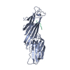
| ||||||||
|---|---|---|---|---|---|---|---|---|---|
| 1 |
| ||||||||
| Unit cell |
|
-Components
| #1: Protein | Mass: 32758.363 Da / Num. of mol.: 1 / Fragment: INTERNALIZATION SIGNAL BINDING DOMAIN Source method: isolated from a genetically manipulated source Details: THE FIRST SEVEN RESIDUES OF THE POLYMER MAKE UP A HIS-TAG Source: (gene. exp.) Rattus norvegicus (Norway rat) / Plasmid: PMW172H6 / Species (production host): Escherichia coli / Gene (production host): AP50 / Production host:  Escherichia coli BL21 (bacteria) / Strain (production host): BL21 / References: UniProt: P84092 Escherichia coli BL21 (bacteria) / Strain (production host): BL21 / References: UniProt: P84092 |
|---|---|
| #2: Protein/peptide | Mass: 808.860 Da / Num. of mol.: 1 / Source method: obtained synthetically Details: HEXAPEPTIDE INTERNALISATION SIGNAL MOTIF FROM TGN38 DYQRLN Source: (synth.) synthetic construct (others) |
| #3: Water | ChemComp-HOH / Water Mass: 18.015 Da / Num. of mol.: 49 / Source method: isolated from a natural source / Formula: H2O Mass: 18.015 Da / Num. of mol.: 49 / Source method: isolated from a natural source / Formula: H2O |
-Experimental details
-Experiment
| Experiment | Method: X-RAY DIFFRACTION / Number of used crystals: 1 |
|---|
- Sample preparation
| Crystal | Density Matthews: 4.98 Å3/Da / Density % sol: 75.3 % / Description: ISOMORPHOUS TO 1BW8 | ||||||||||||||||||||||||||||||||||||
|---|---|---|---|---|---|---|---|---|---|---|---|---|---|---|---|---|---|---|---|---|---|---|---|---|---|---|---|---|---|---|---|---|---|---|---|---|---|
| Crystal grow | Temperature: 289 K / Method: vapor diffusion, hanging drop / pH: 7.1 Details: HANGING DROP, 2.2M NACL, 0.4M NA/K PHOSPHATE, 10MM DTT 0.1M MES PH 7.1, 15% GLYCEROL, 16DEGREES, MOLAR RATIO OF PEPTIDE TO PROTEIN 3:1, vapor diffusion - hanging drop, temperature 289K | ||||||||||||||||||||||||||||||||||||
| Crystal grow | *PLUS Temperature: 16 ℃ / Method: vapor diffusion, hanging drop / PH range low: 7.1 / PH range high: 6.5 | ||||||||||||||||||||||||||||||||||||
| Components of the solutions | *PLUS
|
-Data collection
| Diffraction | Mean temperature: 100 K |
|---|---|
| Diffraction source | Source: SYNCHROTRON / Site: SRS  / Beamline: PX9.6 / Wavelength: 0.877 / Beamline: PX9.6 / Wavelength: 0.877 |
| Detector | Type: ADSC / Detector: CCD / Date: Sep 12, 1998 / Details: MIRROR |
| Radiation | Monochromator: YES / Protocol: SINGLE WAVELENGTH / Monochromatic (M) / Laue (L): M / Scattering type: x-ray |
| Radiation wavelength | Wavelength: 0.877 Å / Relative weight: 1 |
| Reflection | Resolution: 2.7→22 Å / Num. obs: 18413 / % possible obs: 99.8 % / Observed criterion σ(I): 4 / Redundancy: 15.8 % / Biso Wilson estimate: 78 Å2 / Rmerge(I) obs: 0.101 / Rsym value: 0.101 / Net I/σ(I): 23.5 |
| Reflection shell | Resolution: 2.7→2.85 Å / Redundancy: 14.7 % / Rmerge(I) obs: 0.9999999 / Mean I/σ(I) obs: 2.2 / Rsym value: 0.99999 / % possible all: 99.8 |
| Reflection shell | *PLUS % possible obs: 99.8 % |
- Processing
| Software |
| ||||||||||||||||||||||||||||||||||||||||||||||||||||||||||||||||||||||||||||||||||||
|---|---|---|---|---|---|---|---|---|---|---|---|---|---|---|---|---|---|---|---|---|---|---|---|---|---|---|---|---|---|---|---|---|---|---|---|---|---|---|---|---|---|---|---|---|---|---|---|---|---|---|---|---|---|---|---|---|---|---|---|---|---|---|---|---|---|---|---|---|---|---|---|---|---|---|---|---|---|---|---|---|---|---|---|---|---|
| Refinement | Method to determine structure: MOLECULAR REPLACEMENT Starting model: 1BW8 Resolution: 2.7→22 Å / Cross valid method: THROUGHOUT / σ(F): 0 / ESU R Free: 0.32
| ||||||||||||||||||||||||||||||||||||||||||||||||||||||||||||||||||||||||||||||||||||
| Displacement parameters | Biso mean: 78 Å2 | ||||||||||||||||||||||||||||||||||||||||||||||||||||||||||||||||||||||||||||||||||||
| Refinement step | Cycle: LAST / Resolution: 2.7→22 Å
| ||||||||||||||||||||||||||||||||||||||||||||||||||||||||||||||||||||||||||||||||||||
| Refine LS restraints |
|
