 Movie
Movie Controller


+ Open data

- Basic information
| Entry | Database: PDB / ID: 3qdh | ||||||
|---|---|---|---|---|---|---|---|
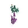
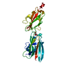
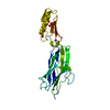
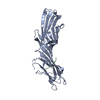
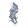
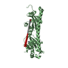
| Title | Crystal structure of Actinomyces fimbrial adhesin FimA | ||||||
 Components Components | Fimbrial structural subunit | ||||||
 Keywords Keywords |  CELL ADHESION / isopeptide bonds / Actinomyces type 2 fimbriae / CnaA/DEv-IgG fold / CnaB/IgG-rev fold / Gram-positive bacterial cell wall protein / fimbrial structural subunit / CELL AHDESION / pilin CELL ADHESION / isopeptide bonds / Actinomyces type 2 fimbriae / CnaA/DEv-IgG fold / CnaB/IgG-rev fold / Gram-positive bacterial cell wall protein / fimbrial structural subunit / CELL AHDESION / pilin | ||||||
| Function / homology |  Function and homology information Function and homology information | ||||||
| Biological species |  Actinomyces naeslundii (bacteria) Actinomyces naeslundii (bacteria) | ||||||
| Method | X-RAY DIFFRACTION / SYNCHROTRON / MIRAS / Resolution: 1.9 Å | ||||||
 Authors Authors | Devarajan, B. / Krishnan, V. / Narayana, S.V.L. | ||||||
 Citation Citation | Journal: Mol.Microbiol. / Year: 2011 Title: Two autonomous structural modules in the fimbrial shaft adhesin FimA mediate Actinomyces interactions with streptococci and host cells during oral biofilm development. Authors: Mishra, A. / Devarajan, B. / Reardon, M.E. / Dwivedi, P. / Krishnan, V. / Cisar, J.O. / Das, A. / Narayana, S.V. / Ton-That, H. | ||||||
| History |
|
- Structure visualization
| Structure viewer | Molecule: MolmilJmol/JSmol |
|---|
- Downloads & links
-Download
| PDBx/mmCIF format | 3qdh.cif.gz | 63.7 KB | Display | PDBx/mmCIF format |
|---|---|---|---|---|
| PDB format | pdb3qdh.ent.gz | 50.2 KB | Display | PDB format |
| PDBx/mmJSON format | 3qdh.json.gz | Tree view | PDBx/mmJSON format | |
| Others |  Other downloads Other downloads |
-Validation report
| Arichive directory | https://data.pdbj.org/pub/pdb/validation_reports/qd/3qdhftp://data.pdbj.org/pub/pdb/validation_reports/qd/3qdh | HTTPS FTP |
|---|
-Related structure data
| Similar structure data |
|---|


-Links
 PDBj
PDBj- Assembly
| Deposited unit | 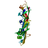
| ||||||||
|---|---|---|---|---|---|---|---|---|---|
| 1 |
| ||||||||
| Unit cell |
| ||||||||
| Details | monomer |
-Components
| #1: Protein | Mass: 31017.447 Da / Num. of mol.: 1 / Fragment: C-terminal domains, residues 199-488 Source method: isolated from a genetically manipulated source Source: (gene. exp.) Actinomyces naeslundii (bacteria) / Strain: T14V / Gene: fimA / Plasmid: pMCSG7 / Production host: Escherichia coli (E. coli) / Strain (production host): BL21(DE3) / References: UniProt: O68212 |
|---|---|
| #2: Chemical | ChemComp-ZN /   Mass: 65.409 Da / Num. of mol.: 1 / Source method: obtained synthetically / Formula: Zn Mass: 65.409 Da / Num. of mol.: 1 / Source method: obtained synthetically / Formula: Zn |
| #3: Water | ChemComp-HOH / Water Mass: 18.015 Da / Num. of mol.: 199 / Source method: isolated from a natural source / Formula: H2O Mass: 18.015 Da / Num. of mol.: 199 / Source method: isolated from a natural source / Formula: H2O |
-Experimental details
-Experiment
| Experiment | Method: X-RAY DIFFRACTION / Number of used crystals: 1 |
|---|
- Sample preparation
| Crystal | Density Matthews: 2.33 Å3/Da / Density % sol: 47.15 % |
|---|---|
| Crystal grow | Temperature: 295 K / Method: vapor diffusion, hanging drop / pH: 8 Details: 17% PEG 2000 MME, 0.1M imidazole, 0.2M zinc acetate, pH 8.0, VAPOR DIFFUSION, HANGING DROP, temperature 295K |
-Data collection
| Diffraction |
| ||||||||||||||||||
|---|---|---|---|---|---|---|---|---|---|---|---|---|---|---|---|---|---|---|---|
| Diffraction source |
| ||||||||||||||||||
| Detector |
| ||||||||||||||||||
| Radiation |
| ||||||||||||||||||
| Radiation wavelength |
| ||||||||||||||||||
| Reflection | Resolution: 1.9→40 Å / Num. obs: 22677 / % possible obs: 99.3 % / Redundancy: 3.5 % / Rmerge(I) obs: 0.063 | ||||||||||||||||||
| Reflection shell | Resolution: 1.9→1.97 Å / Redundancy: 3.5 % / Rmerge(I) obs: 0.203 / Mean I/σ(I) obs: 11.8 / % possible all: 99.5 |

- Processing
| Software |
| |||||||||||||||||||||||||||||||||||||||||||||||||||||||||||||||||
|---|---|---|---|---|---|---|---|---|---|---|---|---|---|---|---|---|---|---|---|---|---|---|---|---|---|---|---|---|---|---|---|---|---|---|---|---|---|---|---|---|---|---|---|---|---|---|---|---|---|---|---|---|---|---|---|---|---|---|---|---|---|---|---|---|---|---|
| Refinement | Method to determine structure: MIRAS / Resolution: 1.9→40 Å / Cor.coef. Fo:Fc: 0.941 / Cor.coef. Fo:Fc free: 0.922 / SU B: 3.709 / SU ML: 0.111 / Cross valid method: THROUGHOUT / ESU R Free: 0.156 / Stereochemistry target values: MAXIMUM LIKELIHOOD / Details: HYDROGENS HAVE BEEN ADDED IN THE RIDING POSITIONS
| |||||||||||||||||||||||||||||||||||||||||||||||||||||||||||||||||
| Solvent computation | Ion probe radii: 0.8 Å / Shrinkage radii: 0.8 Å / VDW probe radii: 1.4 Å / Solvent model: MASK | |||||||||||||||||||||||||||||||||||||||||||||||||||||||||||||||||
| Displacement parameters | Biso mean: 28.707 Å2
| |||||||||||||||||||||||||||||||||||||||||||||||||||||||||||||||||
| Refinement step | Cycle: LAST / Resolution: 1.9→40 Å
| |||||||||||||||||||||||||||||||||||||||||||||||||||||||||||||||||
| Refine LS restraints |
| |||||||||||||||||||||||||||||||||||||||||||||||||||||||||||||||||
| LS refinement shell | Resolution: 1.9→1.949 Å / Total num. of bins used: 20
|
