 Movie
Movie Controller


+ Open data

- Basic information
| Entry | Database: PDB / ID: 4ncd | ||||||
|---|---|---|---|---|---|---|---|
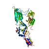
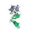
| Title | Crystal Structure of Class 5 Fimbriae Chaperone CfaA | ||||||
 Components Components | Gram-negative pili assembly chaperone, N-terminal domain protein | ||||||
 Keywords Keywords |  CHAPERONE / immunoglobulin fold CHAPERONE / immunoglobulin fold | ||||||
| Function / homology | Immunoglobulin-like - #3970 / Immunoglobulins / Immunoglobulin-like / Sandwich / Mainly Beta / :  Function and homology information Function and homology information | ||||||
| Biological species |  Escherichia coli (E. coli) Escherichia coli (E. coli) | ||||||
| Method | X-RAY DIFFRACTION / SYNCHROTRON / SAD / Resolution: 2.037 Å | ||||||
 Authors Authors | Bao, R. / Xia, D. | ||||||
 Citation Citation | Journal: Plos Pathog. / Year: 2014 Title: Structure of CfaA Suggests a New Family of Chaperones Essential for Assembly of Class 5 Fimbriae. Authors: Bao, R. / Fordyce, A. / Chen, Y.X. / McVeigh, A. / Savarino, S.J. / Xia, D. | ||||||
| History |
|
- Structure visualization
| Structure viewer | Molecule: MolmilJmol/JSmol |
|---|
- Downloads & links
-Download
| PDBx/mmCIF format | 4ncd.cif.gz | 54.5 KB | Display | PDBx/mmCIF format |
|---|---|---|---|---|
| PDB format | pdb4ncd.ent.gz | 41.8 KB | Display | PDB format |
| PDBx/mmJSON format | 4ncd.json.gz | Tree view | PDBx/mmJSON format | |
| Others |  Other downloads Other downloads |
-Validation report
| Arichive directory | https://data.pdbj.org/pub/pdb/validation_reports/nc/4ncdftp://data.pdbj.org/pub/pdb/validation_reports/nc/4ncd | HTTPS FTP |
|---|
-Related structure data
| Similar structure data |
|---|


-Links
 PDBj
PDBj
- Assembly
| Deposited unit | 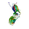
| ||||||||
|---|---|---|---|---|---|---|---|---|---|
| 1 | 
| ||||||||
| 2 |
| ||||||||
| Unit cell |
|
-Components
| #1: Protein | Mass: 28329.703 Da / Num. of mol.: 1 Source method: isolated from a genetically manipulated source Source: (gene. exp.) Escherichia coli (E. coli) / Gene: ECP03018675_4907 / Production host: Escherichia coli (E. coli) / References: UniProt: N4NNE0 |
|---|---|
| #2: Water | ChemComp-HOH / Water Mass: 18.015 Da / Num. of mol.: 120 / Source method: isolated from a natural source / Formula: H2O Mass: 18.015 Da / Num. of mol.: 120 / Source method: isolated from a natural source / Formula: H2O |
-Experimental details
-Experiment
| Experiment | Method: X-RAY DIFFRACTION / Number of used crystals: 1 |
|---|
- Sample preparation
| Crystal | Density Matthews: 2.06 Å3/Da / Density % sol: 40.16 % |
|---|---|
| Crystal grow | Temperature: 293 K / Method: evaporation / pH: 5.3 Details: 22% PEG3350, 0.2 M NaCl, 0.1 M MES pH 5.3, EVAPORATION, temperature 293K |
-Data collection
| Diffraction | Mean temperature: 100 K |
|---|---|
| Diffraction source | Source: SYNCHROTRON / Site: APS  / Beamline: 22-BM / Wavelength: 1.54178 Å / Beamline: 22-BM / Wavelength: 1.54178 Å |
| Detector | Type: MARMOSAIC 225 mm CCD / Detector: CCD / Date: Aug 6, 2010 |
| Radiation | Monochromator: APS BM22 / Protocol: SINGLE WAVELENGTH / Monochromatic (M) / Laue (L): M / Scattering type: x-ray |
| Radiation wavelength | Wavelength: 1.54178 Å / Relative weight: 1 |
| Reflection | Resolution: 2.03→50 Å / Num. obs: 15399 / % possible obs: 97.4 % / Observed criterion σ(F): 0 / Observed criterion σ(I): -1 |
| Reflection shell | Resolution: 2.03→2.08 Å / % possible all: 72.9 |
- Processing
| Software |
| |||||||||||||||||||||||||||||||||||||||||||||||||||||||||||||||||||||||||||||
|---|---|---|---|---|---|---|---|---|---|---|---|---|---|---|---|---|---|---|---|---|---|---|---|---|---|---|---|---|---|---|---|---|---|---|---|---|---|---|---|---|---|---|---|---|---|---|---|---|---|---|---|---|---|---|---|---|---|---|---|---|---|---|---|---|---|---|---|---|---|---|---|---|---|---|---|---|---|---|
| Refinement | Method to determine structure: SAD / Resolution: 2.037→39.715 Å / SU ML: 0.27 / Phase error: 26.48 / Stereochemistry target values: ML
| |||||||||||||||||||||||||||||||||||||||||||||||||||||||||||||||||||||||||||||
| Solvent computation | Shrinkage radii: 0.9 Å / VDW probe radii: 1.11 Å / Solvent model: FLAT BULK SOLVENT MODEL | |||||||||||||||||||||||||||||||||||||||||||||||||||||||||||||||||||||||||||||
| Refinement step | Cycle: LAST / Resolution: 2.037→39.715 Å
| |||||||||||||||||||||||||||||||||||||||||||||||||||||||||||||||||||||||||||||
| Refine LS restraints |
| |||||||||||||||||||||||||||||||||||||||||||||||||||||||||||||||||||||||||||||
| LS refinement shell |
|
