- PDB-4pk6: Crystal structure of the indoleamine 2,3-dioxygenagse 1 (IDO1) co... -
+
Open data
ID or keywords:
Loading...
-
Basic information
Entry
Database: PDB / ID: 4pk6
Title
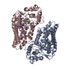
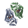
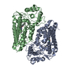
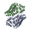
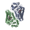
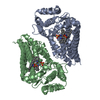
Crystal structure of the indoleamine 2,3-dioxygenagse 1 (IDO1) complexed with imidazothiazole derivative
Components
Indoleamine 2,3-dioxygenase 1
Keywords
OXIDOREDUCTASE/OXIDOREDUCTASE INHIBITOR / indoleamine 2 / 3-dioxygenagse 1 / induced fit / structure based drug discovery / imidazothiazole / OXIDOREDUCTASE-OXIDOREDUCTASE INHIBITOR complex
Function / homology
Function and homology information
indoleamine 2,3-dioxygenase / smooth muscle contractile fiber / indoleamine 2,3-dioxygenase activity / positive regulation of chronic inflammatory response / kynurenic acid biosynthetic process / tryptophan 2,3-dioxygenase activity / positive regulation of T cell tolerance induction / tryptophan catabolic process to kynurenine / stereocilium bundle / positive regulation of type 2 immune response ... indoleamine 2,3-dioxygenase / smooth muscle contractile fiber / indoleamine 2,3-dioxygenase activity / positive regulation of chronic inflammatory response / kynurenic acid biosynthetic process / tryptophan 2,3-dioxygenase activity / positive regulation of T cell tolerance induction / tryptophan catabolic process to kynurenine / stereocilium bundle / positive regulation of type 2 immune response / 'de novo' NAD biosynthetic process from tryptophan / tryptophan catabolic process / Tryptophan catabolism / positive regulation of T cell apoptotic process / negative regulation of T cell apoptotic process / swimming behavior / negative regulation of interleukin-10 production / multicellular organismal response to stress / T cell proliferation / negative regulation of T cell proliferation / positive regulation of interleukin-12 production / female pregnancy / response to lipopolysaccharide / electron transfer activity / inflammatory response / heme binding / metal ion binding / cytosol / cytoplasm Similarity search - Function
Resolution: 3.45→75.65 Å / Cor.coef. Fo:Fc: 0.918 / Cor.coef. Fo:Fc free: 0.877 / SU B: 29.339 / SU ML: 0.458 / Cross valid method: THROUGHOUT / ESU R Free: 0.6 / Stereochemistry target values: MAXIMUM LIKELIHOOD / Details: HYDROGENS HAVE BEEN USED IF PRESENT IN THE INPUT
Rfactor
Num. reflection
% reflection
Selection details
Rfree
0.25604
700
4.9 %
RANDOM
Rwork
0.18999
-
-
-
obs
0.19323
13574
99.99 %
-
Solvent computation
Ion probe radii: 0.8 Å / Shrinkage radii: 0.8 Å / VDW probe radii: 1.2 Å / Solvent model: MASK
 Movie
Movie Controller

 Yorodumi
 Open data
 Basic information
 Components
 Keywords
 Function and homology information
 Authors
 Citation
 Structure visualization
 Downloads & links
 Other downloads
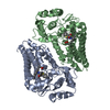


 PDBj
PDBj Assembly
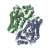


 Mass: 616.487 Da / Num. of mol.: 2 / Source method: obtained synthetically / Formula: C34H32FeN4O4
 Mass: 395.905 Da / Num. of mol.: 2 / Source method: obtained synthetically / Formula: C21H18ClN3OS
 Sample preparation
 / Beamline: X06SA / Wavelength: 1 Å
 Processing