 Movie
Movie Controller

[English] 日本語
 Yorodumi
- PDB-4u72: Crystal structure of 4-phenylimidazole bound form of human indole... -
+ Open data

- Basic information
| Entry | Database: PDB / ID: 4u72 | |||||||||||||||
|---|---|---|---|---|---|---|---|---|---|---|---|---|---|---|---|---|
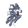
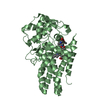
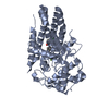
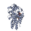
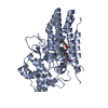
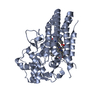
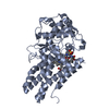
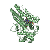
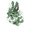
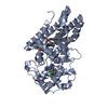
| Title | Crystal structure of 4-phenylimidazole bound form of human indoleamine 2,3-dioxygenase (A260G mutant) | |||||||||||||||
 Components Components | Indoleamine 2,3-dioxygenase 1 | |||||||||||||||
 Keywords Keywords | OXIDOREDUCTASE / metal-binding / all alpha | |||||||||||||||
| Function / homology |  Function and homology information indoleamine 2,3-dioxygenase / smooth muscle contractile fiber / indoleamine 2,3-dioxygenase activity / positive regulation of chronic inflammatory response / kynurenic acid biosynthetic process / tryptophan 2,3-dioxygenase activity / positive regulation of T cell tolerance induction / tryptophan catabolic process to kynurenine / stereocilium bundle / positive regulation of type 2 immune response ... indoleamine 2,3-dioxygenase / smooth muscle contractile fiber / indoleamine 2,3-dioxygenase activity / positive regulation of chronic inflammatory response / kynurenic acid biosynthetic process / tryptophan 2,3-dioxygenase activity / positive regulation of T cell tolerance induction / tryptophan catabolic process to kynurenine / stereocilium bundle / positive regulation of type 2 immune response / 'de novo' NAD biosynthetic process from tryptophan / tryptophan catabolic process / Tryptophan catabolism / positive regulation of T cell apoptotic process / negative regulation of T cell apoptotic process / swimming behavior / negative regulation of interleukin-10 production / multicellular organismal response to stress / T cell proliferation / negative regulation of T cell proliferation / positive regulation of interleukin-12 production / female pregnancy / response to lipopolysaccharide / electron transfer activity / inflammatory response / heme binding / metal ion binding / cytosol / cytoplasm Function and homology information indoleamine 2,3-dioxygenase / smooth muscle contractile fiber / indoleamine 2,3-dioxygenase activity / positive regulation of chronic inflammatory response / kynurenic acid biosynthetic process / tryptophan 2,3-dioxygenase activity / positive regulation of T cell tolerance induction / tryptophan catabolic process to kynurenine / stereocilium bundle / positive regulation of type 2 immune response ... indoleamine 2,3-dioxygenase / smooth muscle contractile fiber / indoleamine 2,3-dioxygenase activity / positive regulation of chronic inflammatory response / kynurenic acid biosynthetic process / tryptophan 2,3-dioxygenase activity / positive regulation of T cell tolerance induction / tryptophan catabolic process to kynurenine / stereocilium bundle / positive regulation of type 2 immune response / 'de novo' NAD biosynthetic process from tryptophan / tryptophan catabolic process / Tryptophan catabolism / positive regulation of T cell apoptotic process / negative regulation of T cell apoptotic process / swimming behavior / negative regulation of interleukin-10 production / multicellular organismal response to stress / T cell proliferation / negative regulation of T cell proliferation / positive regulation of interleukin-12 production / female pregnancy / response to lipopolysaccharide / electron transfer activity / inflammatory response / heme binding / metal ion binding / cytosol / cytoplasmSimilarity search - Function | |||||||||||||||
| Biological species |  Homo sapiens (human) Homo sapiens (human) | |||||||||||||||
| Method | X-RAY DIFFRACTION / SYNCHROTRON / Resolution: 2 Å | |||||||||||||||
 Authors Authors | Sugimoto, H. / Horitani, M. / Kometani, E. / Shiro, Y. | |||||||||||||||
| Funding support |  Japan, 4items Japan, 4items
| |||||||||||||||
 Citation Citation | Journal: to be published Title: Conformation and Mobility of Active Site Loop is Critical for Substrate Binding and Inhibition in Human Indoleamine 2,3-Dioxygenase Authors: Horitani, M. / Kometani, E. / Vottero, E. / Otsuki, T. / Shiro, Y. / Sugimoto, H. #1: Journal: Proc. Natl. Acad. Sci. U.S.A. / Year: 2006Title: Crystal structure of human indoleamine 2,3-dioxygenase: catalytic mechanism of O2 incorporation by a heme-containing dioxygenase. Authors: Sugimoto, H. / Oda, S. / Otsuki, T. / Hino, T. / Yoshida, T. / Shiro, Y. | |||||||||||||||
| History |
|
- Structure visualization
| Structure viewer | Molecule: MolmilJmol/JSmol |
|---|
- Downloads & links
-Download
| PDBx/mmCIF format | 4u72.cif.gz | 168.3 KB | Display | PDBx/mmCIF format |
|---|---|---|---|---|
| PDB format | pdb4u72.ent.gz | 138.1 KB | Display | PDB format |
| PDBx/mmJSON format | 4u72.json.gz | Tree view | PDBx/mmJSON format | |
| Others |  Other downloads Other downloads |
-Validation report
| Arichive directory | https://data.pdbj.org/pub/pdb/validation_reports/u7/4u72ftp://data.pdbj.org/pub/pdb/validation_reports/u7/4u72 | HTTPS FTP |
|---|
-Related structure data


-Links
 PDBj
PDBj- Assembly
| Deposited unit | 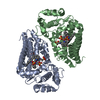
| |||||||||||||||||||||||||||
|---|---|---|---|---|---|---|---|---|---|---|---|---|---|---|---|---|---|---|---|---|---|---|---|---|---|---|---|---|
| 1 | 
| |||||||||||||||||||||||||||
| 2 | 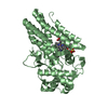
| |||||||||||||||||||||||||||
| Unit cell |
| |||||||||||||||||||||||||||
| Noncrystallographic symmetry (NCS) | NCS domain:
NCS domain segments:
|
-Components
| #1: Protein | / IDO-1 / Indoleamine-pyrrole 2 / 3-dioxygenase Mass: 45652.453 Da / Num. of mol.: 2 / Fragment: indoleamine 2,3-dioxygenase / Mutation: A260G Source method: isolated from a genetically manipulated source Source: (gene. exp.) Homo sapiens (human) / Gene: IDO1, IDO, INDO / Plasmid: pET-15b / Production host:  Escherichia coli (E. coli) / Strain (production host): BL21(DE3) CodonPlus RIL / References: UniProt: P14902, indoleamine 2,3-dioxygenase Escherichia coli (E. coli) / Strain (production host): BL21(DE3) CodonPlus RIL / References: UniProt: P14902, indoleamine 2,3-dioxygenase#2: Chemical | Heme B  Mass: 616.487 Da / Num. of mol.: 2 / Source method: obtained synthetically / Formula: C34H32FeN4O4 Mass: 616.487 Da / Num. of mol.: 2 / Source method: obtained synthetically / Formula: C34H32FeN4O4#3: Chemical |   Mass: 144.173 Da / Num. of mol.: 2 / Source method: obtained synthetically / Formula: C9H8N2 Mass: 144.173 Da / Num. of mol.: 2 / Source method: obtained synthetically / Formula: C9H8N2#4: Chemical | ChemComp-NHE / CHES (buffer)  Mass: 207.290 Da / Num. of mol.: 4 / Source method: obtained synthetically / Formula: C8H17NO3S / Comment: pH buffer*YM Mass: 207.290 Da / Num. of mol.: 4 / Source method: obtained synthetically / Formula: C8H17NO3S / Comment: pH buffer*YM#5: Water | ChemComp-HOH / | Water Mass: 18.015 Da / Num. of mol.: 335 / Source method: isolated from a natural source / Formula: H2O Mass: 18.015 Da / Num. of mol.: 335 / Source method: isolated from a natural source / Formula: H2O |
|---|
-Experimental details
-Experiment
| Experiment | Method: X-RAY DIFFRACTION / Number of used crystals: 1 |
|---|
- Sample preparation
| Crystal | Density Matthews: 3.06 Å3/Da / Density % sol: 59.84 % |
|---|---|
| Crystal grow | Temperature: 293 K / Method: vapor diffusion, hanging drop / pH: 9.1 Details: 12 % (W/V) PEG 8000, 0.2 M ammonium acetate, 1 mM 4-phenylimidazole, 0.1 M CHES |
-Data collection
| Diffraction | Mean temperature: 100 K |
|---|---|
| Diffraction source | Source: SYNCHROTRON / Site: SPring-8 / Beamline: BL26B1 / Wavelength: 1 Å |
| Detector | Type: RIGAKU JUPITER 210 / Detector: CCD / Date: Feb 7, 2009 / Details: mirrors |
| Diffraction measurement | Details: 0.60 degrees, 10.0 sec, detector distance 169.70 mm Method: \w scans |
| Radiation | Monochromator: Si(111) / Protocol: SINGLE WAVELENGTH / Monochromatic (M) / Laue (L): M / Scattering type: x-ray |
| Radiation wavelength | Wavelength: 1 Å / Relative weight: 1 |
| Reflection | Av R equivalents: 0.054 / Number: 581251 |
| Reflection | Resolution: 2→40 Å / Num. obs: 76205 / % possible obs: 99.4 % / Observed criterion σ(F): 0 / Observed criterion σ(I): -3 / Redundancy: 7.6 % / Biso Wilson estimate: 29.1 Å2 / Rmerge(I) obs: 0.054 / Rsym value: 0.054 / Net I/σ(I): 37.033 |
| Reflection shell | Resolution: 2→2.07 Å / Redundancy: 5.9 % / Rmerge(I) obs: 0.464 / Mean I/σ(I) obs: 2.75 / Rsym value: 0.464 / % possible all: 94.9 |
| Cell measurement | Reflection used: 581251 |
- Processing
| Software |
| |||||||||||||||||||||||||||||||||||||||||||||
|---|---|---|---|---|---|---|---|---|---|---|---|---|---|---|---|---|---|---|---|---|---|---|---|---|---|---|---|---|---|---|---|---|---|---|---|---|---|---|---|---|---|---|---|---|---|---|
| Refinement | Resolution: 2→19.76 Å / Cor.coef. Fo:Fc: 0.955 / Cor.coef. Fo:Fc free: 0.931 / WRfactor Rfree: 0.2156 / WRfactor Rwork: 0.1744 / FOM work R set: 0.842 / SU B: 3.474 / SU ML: 0.097 / SU R Cruickshank DPI: 0.1453 / SU Rfree: 0.1439 / Cross valid method: THROUGHOUT / σ(F): 0 / ESU R: 0.145 / ESU R Free: 0.144 / Stereochemistry target values: MAXIMUM LIKELIHOOD Details: HYDROGENS HAVE BEEN USED IF PRESENT IN THE INPUT U VALUES
| |||||||||||||||||||||||||||||||||||||||||||||
| Solvent computation | Ion probe radii: 0.8 Å / Shrinkage radii: 0.8 Å / VDW probe radii: 1.2 Å / Solvent model: MASK | |||||||||||||||||||||||||||||||||||||||||||||
| Displacement parameters | Biso max: 126.77 Å2 / Biso mean: 27.393 Å2 / Biso min: 11.61 Å2
| |||||||||||||||||||||||||||||||||||||||||||||
| Refinement step | Cycle: final / Resolution: 2→19.76 Å
| |||||||||||||||||||||||||||||||||||||||||||||
| Refine LS restraints |
| |||||||||||||||||||||||||||||||||||||||||||||
| Refine LS restraints NCS | Ens-ID: 1 / Number: 477 / Refine-ID: X-RAY DIFFRACTION / Type: interatomic distance / Rms dev position: 0.12 Å / Weight position: 0.05
| |||||||||||||||||||||||||||||||||||||||||||||
| LS refinement shell | Resolution: 1.997→2.048 Å / Total num. of bins used: 20
|
