[English] 日本語
Yorodumi
- PDB-6ubp: Crystal structure of a photochemical intermediate of human indole... -

+
Open data


ID or keywords:

Loading...

-
Basic information

Entry
Database: PDB / ID: 6ubp
Title
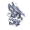
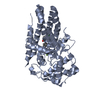
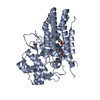
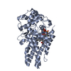
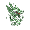
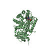
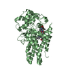
Crystal structure of a photochemical intermediate of human indoleamine 2,3-dioxygenase 1 in complex with carbon monoxide and tryptophan
ComponentsIndoleamine 2,3-dioxygenase 1
KeywordsOXIDOREDUCTASE / human Indoleamine 2 / 3-dioxygenase x-ray photolysis of CO and Tryptophan
Function / homology
Function and homology information


indoleamine 2,3-dioxygenase / smooth muscle contractile fiber / indoleamine 2,3-dioxygenase activity / positive regulation of chronic inflammatory response / kynurenic acid biosynthetic process / tryptophan 2,3-dioxygenase activity / positive regulation of T cell tolerance induction / tryptophan catabolic process to kynurenine / stereocilium bundle / positive regulation of type 2 immune response ... indoleamine 2,3-dioxygenase / smooth muscle contractile fiber / indoleamine 2,3-dioxygenase activity / positive regulation of chronic inflammatory response / kynurenic acid biosynthetic process / tryptophan 2,3-dioxygenase activity / positive regulation of T cell tolerance induction / tryptophan catabolic process to kynurenine / stereocilium bundle / positive regulation of type 2 immune response / 'de novo' NAD biosynthetic process from tryptophan / tryptophan catabolic process / Tryptophan catabolism / positive regulation of T cell apoptotic process / negative regulation of T cell apoptotic process / swimming behavior / negative regulation of interleukin-10 production / multicellular organismal response to stress / T cell proliferation / negative regulation of T cell proliferation / positive regulation of interleukin-12 production / female pregnancy / response to lipopolysaccharide / electron transfer activity / inflammatory response / heme binding / metal ion binding / cytosol / cytoplasm
Similarity search - Function
Indoleamine 2,3-dioxygenase / Indoleamine 2,3-dioxygenase / Indoleamine 2,3-dioxygenase signature 1. / Indoleamine 2,3-dioxygenase signature 2. / Tryptophan/Indoleamine 2,3-dioxygenase-like
Similarity search - Domain/homology
CARBON MONOXIDE / PROTOPORPHYRIN IX CONTAINING FE / TRYPTOPHAN / Indoleamine 2,3-dioxygenase 1
Similarity search - Component
Biological speciesHomo sapiens (human)
MethodX-RAY DIFFRACTION / SYNCHROTRON / MOLECULAR REPLACEMENT / Resolution: 2.95 Å
AuthorsPham, K.N. / Yeh, S.R.
Funding support United States, 2items
OrganizationGrant numberCountry
National Institutes of Health/National Institute of General Medical Sciences (NIH/NIGMS)GM115773 United States
National Science Foundation (NSF, United States)CHE-1404929 United States
CitationJournal: J.Am.Chem.Soc. / Year: 2021
Title: Conformational Plasticity in Human Heme-Based Dioxygenases.
Authors: Pham, K.N. / Lewis-Ballester, A. / Yeh, S.R.
History
DepositionSep 12, 2019Deposition site: RCSB / Processing site: RCSB
Revision 1.0Feb 3, 2021Provider: repository / Type: Initial release
Revision 1.1Feb 17, 2021Group: Database references / Category: citation / citation_author
Item: _citation.journal_volume / _citation.page_first ..._citation.journal_volume / _citation.page_first / _citation.page_last / _citation.year / _citation_author.identifier_ORCID
Revision 1.2Oct 11, 2023Group: Data collection / Database references / Refinement description
Category: chem_comp_atom / chem_comp_bond ...chem_comp_atom / chem_comp_bond / database_2 / pdbx_initial_refinement_model
Item: _database_2.pdbx_DOI / _database_2.pdbx_database_accession

-
Structure visualization

Structure viewerMolecule:
MolmilJmol/JSmol

Downloads & links

-
Assembly

Deposited unit
A: Indoleamine 2,3-dioxygenase 1
B: Indoleamine 2,3-dioxygenase 1
hetero molecules


Theoretical massNumber of molelcules
Total (without water)97,2818
Polymers95,5842
Non-polymers1,6976
Water1,13563
1
A: Indoleamine 2,3-dioxygenase 1
hetero molecules


Theoretical massNumber of molelcules
Total (without water)48,6414
Polymers47,7921
Non-polymers8493
Water181
TypeNameSymmetry operationNumber
identity operation1_555x,y,z1
2
B: Indoleamine 2,3-dioxygenase 1
hetero molecules


Theoretical massNumber of molelcules
Total (without water)48,6414
Polymers47,7921
Non-polymers8493
Water181
TypeNameSymmetry operationNumber
identity operation1_555x,y,z1
Unit cell
Length a, b, c (Å)86.944, 97.727, 130.628
Angle α, β, γ (deg.)90.000, 90.000, 90.000
Int Tables number19
Space group name H-MP212121

-
Components

#1: Protein Indoleamine 2,3-dioxygenase 1 / / IDO-1 / Indoleamine-pyrrole 2 / 3-dioxygenase


Mass: 47791.918 Da / Num. of mol.: 2
Source method: isolated from a genetically manipulated source
Source: (gene. exp.) Homo sapiens (human) / Gene: IDO1, IDO, INDO / Production host: Escherichia coli (E. coli) / References: UniProt: P14902, indoleamine 2,3-dioxygenase
#2: Chemical ChemComp-HEM / PROTOPORPHYRIN IX CONTAINING FE / HEME / Heme B


Mass: 616.487 Da / Num. of mol.: 2 / Source method: obtained synthetically / Formula: C34H32FeN4O4 / Feature type: SUBJECT OF INVESTIGATION
#3: Chemical ChemComp-TRP / TRYPTOPHAN / Tryptophan


Type: L-peptide linking / Mass: 204.225 Da / Num. of mol.: 2 / Source method: obtained synthetically / Formula: C11H12N2O2 / Feature type: SUBJECT OF INVESTIGATION
#4: Chemical ChemComp-CMO / CARBON MONOXIDE / Carbon monoxide


Mass: 28.010 Da / Num. of mol.: 2 / Source method: obtained synthetically / Formula: CO / Feature type: SUBJECT OF INVESTIGATION
#5: Water ChemComp-HOH / water / Water


Mass: 18.015 Da / Num. of mol.: 63 / Source method: isolated from a natural source / Formula: H2O
Has ligand of interestY

-
Experimental details

-
Experiment

ExperimentMethod: X-RAY DIFFRACTION / Number of used crystals: 1

-
Sample preparation

CrystalDensity Matthews: 2.9 Å3/Da / Density % sol: 57.63 %
Crystal growTemperature: 277 K / Method: microbatch / pH: 10
Details: 100 mM sodium thiosulfate, 100 mM CAPS, pH 10.0, 20% w/v PEG8000

-
Data collection

DiffractionMean temperature: 100 K / Serial crystal experiment: N
Diffraction sourceSource: SYNCHROTRON / Site: NSLS-II / Beamline: 17-ID-2 / Wavelength: 0.97931 Å
DetectorType: DECTRIS EIGER X 16M / Detector: PIXEL / Date: May 25, 2019
RadiationMonochromator: double crystal Si(111) / Protocol: SINGLE WAVELENGTH / Monochromatic (M) / Laue (L): M / Scattering type: x-ray
Radiation wavelengthWavelength: 0.97931 Å / Relative weight: 1
ReflectionResolution: 2.95→29.34 Å / Num. obs: 24106 / % possible obs: 99.9 % / Redundancy: 7.6 % / CC1/2: 1 / Rmerge(I) obs: 0.098 / Net I/σ(I): 12.2
Reflection shellResolution: 2.95→3.13 Å / Redundancy: 8 % / Rmerge(I) obs: 2.015 / Mean I/σ(I) obs: 1.2 / Num. unique obs: 3854 / CC1/2: 0.53 / % possible all: 100

-
Processing

Software
NameVersionClassification
REFMAC5.8.0238refinement
PDB_EXTRACT3.25data extraction
XDSdata reduction
Aimlessdata scaling
PHASERphasing
RefinementMethod to determine structure: MOLECULAR REPLACEMENT
Starting model: PDB entry 5WMU
Resolution: 2.95→29.34 Å / Cor.coef. Fo:Fc: 0.958 / Cor.coef. Fo:Fc free: 0.932 / SU B: 22.175 / SU ML: 0.377 / Cross valid method: THROUGHOUT / σ(F): 0 / ESU R Free: 0.41 / Stereochemistry target values: MAXIMUM LIKELIHOOD
Details: HYDROGENS HAVE BEEN ADDED IN THE RIDING POSITIONS U VALUES : REFINED INDIVIDUALLY
RfactorNum. reflection% reflectionSelection details
Rfree0.2641 1165 4.8 %RANDOM
Rwork0.2152 ---
obs0.2175 22893 99.85 %-
Solvent computationIon probe radii: 0.8 Å / Shrinkage radii: 0.8 Å / VDW probe radii: 1.2 Å / Solvent model: MASK
Displacement parametersBiso max: 171.05 Å2 / Biso mean: 102.551 Å2 / Biso min: 67.1 Å2
Baniso -1Baniso -2Baniso -3
1--1.87 Å20 Å2-0 Å2
2---3.07 Å20 Å2
3---4.94 Å2
Refinement stepCycle: final / Resolution: 2.95→29.34 Å
ProteinNucleic acidLigandSolventTotal
Num. atoms5932 0 120 63 6115
Biso mean--108.32 87.61 -
Num. residues----748
Refine LS restraints
Refine-IDTypeDev idealDev ideal targetNumber
X-RAY DIFFRACTIONr_bond_refined_d0.0020.0136205
X-RAY DIFFRACTIONr_bond_other_d0.0010.0175798
X-RAY DIFFRACTIONr_angle_refined_deg1.2291.6618420
X-RAY DIFFRACTIONr_angle_other_deg1.0341.58913455
X-RAY DIFFRACTIONr_dihedral_angle_1_deg5.8135744
X-RAY DIFFRACTIONr_dihedral_angle_2_deg32.74722.656305
X-RAY DIFFRACTIONr_dihedral_angle_3_deg18.455151073
X-RAY DIFFRACTIONr_dihedral_angle_4_deg14.0621532
X-RAY DIFFRACTIONr_chiral_restr0.0410.2761
X-RAY DIFFRACTIONr_gen_planes_refined0.0030.026872
X-RAY DIFFRACTIONr_gen_planes_other0.0030.021308
LS refinement shellResolution: 2.95→3.026 Å / Rfactor Rfree error: 0 / Total num. of bins used: 20
RfactorNum. reflection% reflection
Rfree0.423 87 -
Rwork0.373 1670 -
all-1757 -
obs--100 %

+
About Yorodumi

-
News

-
Feb 9, 2022. New format data for meta-information of EMDB entries

New format data for meta-information of EMDB entries

  • Version 3 of the EMDB header file is now the official format.
  • The previous official version 1.9 will be removed from the archive.

Related info.:EMDB header

External links:wwPDB to switch to version 3 of the EMDB data model

-
Aug 12, 2020. Covid-19 info

Covid-19 info

URL: https://pdbj.org/emnavi/covid19.php

New page: Covid-19 featured information page in EM Navigator.

Related info.:Covid-19 info / Mar 5, 2020. Novel coronavirus structure data

+
Mar 5, 2020. Novel coronavirus structure data

Novel coronavirus structure data

Related info.:Yorodumi Speices / Aug 12, 2020. Covid-19 info

External links:COVID-19 featured content - PDBj / Molecule of the Month (242):Coronavirus Proteases

+
Jan 31, 2019. EMDB accession codes are about to change! (news from PDBe EMDB page)

EMDB accession codes are about to change! (news from PDBe EMDB page)

  • The allocation of 4 digits for EMDB accession codes will soon come to an end. Whilst these codes will remain in use, new EMDB accession codes will include an additional digit and will expand incrementally as the available range of codes is exhausted. The current 4-digit format prefixed with “EMD-” (i.e. EMD-XXXX) will advance to a 5-digit format (i.e. EMD-XXXXX), and so on. It is currently estimated that the 4-digit codes will be depleted around Spring 2019, at which point the 5-digit format will come into force.
  • The EM Navigator/Yorodumi systems omit the EMD- prefix.

Related info.:Q: What is EMD? / ID/Accession-code notation in Yorodumi/EM Navigator

External links:EMDB Accession Codes are Changing Soon! / Contact to PDBj

+
Jul 12, 2017. Major update of PDB

Major update of PDB

  • wwPDB released updated PDB data conforming to the new PDBx/mmCIF dictionary.
  • This is a major update changing the version number from 4 to 5, and with Remediation, in which all the entries are updated.
  • In this update, many items about electron microscopy experimental information are reorganized (e.g. em_software).
  • Now, EM Navigator and Yorodumi are based on the updated data.

External links:wwPDB Remediation / Enriched Model Files Conforming to OneDep Data Standards Now Available in the PDB FTP Archive

-
Yorodumi

Thousand views of thousand structures

  • Yorodumi is a browser for structure data from EMDB, PDB, SASBDB, etc.
  • This page is also the successor to EM Navigator detail page, and also detail information page/front-end page for Omokage search.
  • The word "yorodu" (or yorozu) is an old Japanese word meaning "ten thousand". "mi" (miru) is to see.

Related info.:EMDB / PDB / SASBDB / Comparison of 3 databanks / Yorodumi Search / Aug 31, 2016. New EM Navigator & Yorodumi / Yorodumi Papers / Jmol/JSmol / Function and homology information / Changes in new EM Navigator and Yorodumi

Read more