- PDB-6f0a: Crystal structure of human indoleamine 2,3-dioxygenase bound to a... -
+
Open data
ID or keywords:
Loading...
-
Basic information
Entry
Database: PDB / ID: 6f0a
Title
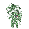
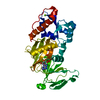
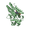
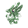
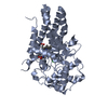
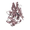
Crystal structure of human indoleamine 2,3-dioxygenase bound to a triazole inhibitor and alanine molecule.
Components
Indoleamine 2,3-dioxygenase 1
Keywords
OXIDOREDUCTASE / Tryptophan 2 3 Dioxygenase Activity Electron Transfer Activity Oxidoreductase Activity Heme Binding Indoleamine 2 3 Dioxygenase Activity Metal Ion Binding Dioxygenase Activity
Function / homology
Function and homology information
indoleamine 2,3-dioxygenase / smooth muscle contractile fiber / indoleamine 2,3-dioxygenase activity / positive regulation of chronic inflammatory response / kynurenic acid biosynthetic process / tryptophan 2,3-dioxygenase activity / positive regulation of T cell tolerance induction / tryptophan catabolic process to kynurenine / stereocilium bundle / positive regulation of type 2 immune response ... indoleamine 2,3-dioxygenase / smooth muscle contractile fiber / indoleamine 2,3-dioxygenase activity / positive regulation of chronic inflammatory response / kynurenic acid biosynthetic process / tryptophan 2,3-dioxygenase activity / positive regulation of T cell tolerance induction / tryptophan catabolic process to kynurenine / stereocilium bundle / positive regulation of type 2 immune response / 'de novo' NAD biosynthetic process from tryptophan / tryptophan catabolic process / Tryptophan catabolism / positive regulation of T cell apoptotic process / negative regulation of T cell apoptotic process / swimming behavior / negative regulation of interleukin-10 production / multicellular organismal response to stress / T cell proliferation / negative regulation of T cell proliferation / positive regulation of interleukin-12 production / female pregnancy / response to lipopolysaccharide / electron transfer activity / inflammatory response / heme binding / metal ion binding / cytosol / cytoplasm Similarity search - Function
In the structure databanks used in Yorodumi, some data are registered as the other names, "COVID-19 virus" and "2019-nCoV". Here are the details of the virus and the list of structure data.
Jan 31, 2019. EMDB accession codes are about to change! (news from PDBe EMDB page)
EMDB accession codes are about to change! (news from PDBe EMDB page)
The allocation of 4 digits for EMDB accession codes will soon come to an end. Whilst these codes will remain in use, new EMDB accession codes will include an additional digit and will expand incrementally as the available range of codes is exhausted. The current 4-digit format prefixed with “EMD-” (i.e. EMD-XXXX) will advance to a 5-digit format (i.e. EMD-XXXXX), and so on. It is currently estimated that the 4-digit codes will be depleted around Spring 2019, at which point the 5-digit format will come into force.
The EM Navigator/Yorodumi systems omit the EMD- prefix.
Related info.:Q: What is EMD? / ID/Accession-code notation in Yorodumi/EM Navigator
Yorodumi is a browser for structure data from EMDB, PDB, SASBDB, etc.
This page is also the successor to EM Navigator detail page, and also detail information page/front-end page for Omokage search.
The word "yorodu" (or yorozu) is an old Japanese word meaning "ten thousand". "mi" (miru) is to see.
Related info.:EMDB / PDB / SASBDB / Comparison of 3 databanks / Yorodumi Search / Aug 31, 2016. New EM Navigator & Yorodumi / Yorodumi Papers / Jmol/JSmol / Function and homology information / Changes in new EM Navigator and Yorodumi
 Movie
Movie Controller

 Yorodumi
 Open data
 Basic information
 Components
 Keywords
 Function and homology information
 Authors
 Citation
 Structure visualization
 Downloads & links
 Other downloads
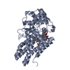


 PDBj
PDBj Assembly
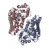


 Mass: 194.621 Da / Num. of mol.: 2 / Source method: obtained synthetically / Formula: C8H7ClN4 / Feature type: SUBJECT OF INVESTIGATION
 Type: L-peptide linking / Mass: 89.093 Da / Num. of mol.: 2 / Source method: obtained synthetically / Formula: C3H7NO2
 Mass: 616.487 Da / Num. of mol.: 2 / Source method: obtained synthetically / Formula: C34H32FeN4O4
 Mass: 18.015 Da / Num. of mol.: 217 / Source method: isolated from a natural source / Formula: H2O
 Sample preparation
 / Beamline: I03 / Wavelength: 0.9763 Å
 Processing