 Movie
Movie Controller


+ Open data

- Basic information
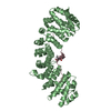
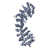
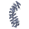
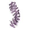
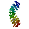
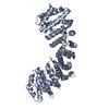
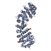
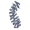
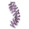
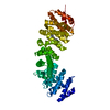
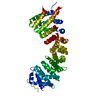
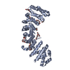
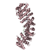
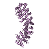
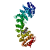
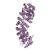
| Entry | Database: PDB / ID: 4mz5 | |||||||||
|---|---|---|---|---|---|---|---|---|---|---|
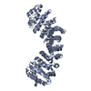
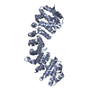
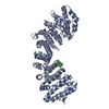
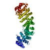
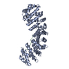
| Title | Structure of importin-alpha: dUTPase NLS complex | |||||||||
 Components Components |
| |||||||||
 Keywords Keywords |  PROTEIN TRANSPORT / ARM repeat / Importin / nucleus PROTEIN TRANSPORT / ARM repeat / Importin / nucleus | |||||||||
| Function / homology |  Function and homology information Function and homology informationSensing of DNA Double Strand Breaks / dUTP catabolic process / entry of viral genome into host nucleus through nuclear pore complex via importin / dUMP biosynthetic process / positive regulation of viral life cycle / dUTP diphosphatase / dUTP diphosphatase activity / NLS-dependent protein nuclear import complex / postsynapse to nucleus signaling pathway / Interconversion of nucleotide di- and triphosphates ...Sensing of DNA Double Strand Breaks / dUTP catabolic process / entry of viral genome into host nucleus through nuclear pore complex via importin / dUMP biosynthetic process / positive regulation of viral life cycle / dUTP diphosphatase / dUTP diphosphatase activity / NLS-dependent protein nuclear import complex / postsynapse to nucleus signaling pathway / Interconversion of nucleotide di- and triphosphates / nucleobase-containing compound metabolic process / nuclear import signal receptor activity / nuclear localization sequence binding / dTMP biosynthetic process / NLS-bearing protein import into nucleus / host cell / cytoplasmic stress granule / protein import into nucleus / DNA-binding transcription factor binding / DNA replication / postsynaptic density / glutamatergic synapse / magnesium ion binding / mitochondrion / RNA binding / extracellular exosome / nucleoplasm / nucleus / cytosolSimilarity search - Function | |||||||||
| Biological species |  Mus musculus (house mouse) Mus musculus (house mouse) Homo sapiens (human) Homo sapiens (human) | |||||||||
| Method | X-RAY DIFFRACTION / SYNCHROTRON / MOLECULAR REPLACEMENT / Resolution: 2.1 Å | |||||||||
 Authors Authors | Marfori, M. / Rona, G. / Vertessy, B.G. / Kobe, B. | |||||||||
 Citation Citation | Journal: Acta Crystallogr.,Sect.D / Year: 2013 Title: Phosphorylation adjacent to the nuclear localization signal of human dUTPase abolishes nuclear import: structural and mechanistic insights. Authors: Rona, G. / Marfori, M. / Borsos, M. / Scheer, I. / Takacs, E. / Toth, J. / Babos, F. / Magyar, A. / Erdei, A. / Bozoky, Z. / Buday, L. / Kobe, B. / Vertessy, B.G. | |||||||||
| History |
|
- Structure visualization
| Structure viewer | Molecule: MolmilJmol/JSmol |
|---|
- Downloads & links
-Download
| PDBx/mmCIF format | 4mz5.cif.gz | 189.2 KB | Display | PDBx/mmCIF format |
|---|---|---|---|---|
| PDB format | pdb4mz5.ent.gz | 150.6 KB | Display | PDB format |
| PDBx/mmJSON format | 4mz5.json.gz | Tree view | PDBx/mmJSON format | |
| Others |  Other downloads Other downloads |
-Validation report
| Arichive directory | https://data.pdbj.org/pub/pdb/validation_reports/mz/4mz5ftp://data.pdbj.org/pub/pdb/validation_reports/mz/4mz5 | HTTPS FTP |
|---|
-Related structure data


-Links
 PDBj
PDBj


- Assembly
| Deposited unit | 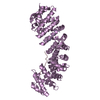
| ||||||||
|---|---|---|---|---|---|---|---|---|---|
| 1 |
| ||||||||
| Unit cell |
| ||||||||
| Details | THE PROTEIN IMPORTIN ALPHA HAS A TWO BINDING SITES ON THE SURFACE OF THE MOLECULE. THERE IS EXTENSIVE LITERATURE THAT SHOWS THAT 'BIPARTITE' PEPTIDES CAN BIND TO BOTH BINDING SITES SIMULTANEOUSLY, WHILE SHORTER 'MONOPARTITE' PEPTIDES SUCH AS THE ONES USED IN THIS STUDY, BIND TO ONLY ONE. WE THEREFORE BELIEVE THAT THE PRESENCE OF THE PEPTIDE AT BOTH BINDING SITES IS THE RESULT OF CRYSTALLIZATION ARTIFACTS DUE TO THE CONCENTRATION OF PEPTIDE USED IN THE STUDY. |
-Components
| #1: Protein/peptide | Mass: 1384.604 Da / Num. of mol.: 2 / Fragment: UNP residues 97-109 / Source method: obtained synthetically / Source: (synth.) Homo sapiens (human) / References: UniProt: P33316#2: Protein | | / Importin alpha P1 / Karyopherin subunit alpha-2 / Pendulin / Pore targeting complex 58 kDa subunit ...Importin alpha P1 / Karyopherin subunit alpha-2 / Pendulin / Pore targeting complex 58 kDa subunit / PTAC58 / RAG cohort protein 1 / SRP1-alphaMass: 55121.301 Da / Num. of mol.: 1 / Fragment: UNP residues 70-528 Source method: isolated from a genetically manipulated source Source: (gene. exp.) Mus musculus (house mouse) / Gene: Kpna2, Rch1 / Production host:  Escherichia coli (E. coli) / Strain (production host): BL21 (DE3) / References: UniProt: P52293 Escherichia coli (E. coli) / Strain (production host): BL21 (DE3) / References: UniProt: P52293#3: Water | ChemComp-HOH / | Water Mass: 18.015 Da / Num. of mol.: 192 / Source method: isolated from a natural source / Formula: H2O Mass: 18.015 Da / Num. of mol.: 192 / Source method: isolated from a natural source / Formula: H2O |
|---|
-Experimental details
-Experiment
| Experiment | Method: X-RAY DIFFRACTION / Number of used crystals: 1 |
|---|
- Sample preparation
| Crystal | Density Matthews: 3.02 Å3/Da / Density % sol: 59.25 % |
|---|---|
| Crystal grow | Temperature: 293 K / Method: vapor diffusion, hanging drop / pH: 7 Details: Sodium citrate, DTT, pH 7, VAPOR DIFFUSION, HANGING DROP, temperature 293K |
-Data collection
| Diffraction | Mean temperature: 100 K |
|---|---|
| Diffraction source | Source: SYNCHROTRON / Site: Australian Synchrotron  / Beamline: MX1 / Wavelength: 0.953693 Å / Beamline: MX1 / Wavelength: 0.953693 Å |
| Detector | Type: ADSC QUANTUM 210r / Detector: CCD / Date: Dec 14, 2011 |
| Radiation | Monochromator: Sagitally focused Si / Protocol: SINGLE WAVELENGTH / Monochromatic (M) / Laue (L): M / Scattering type: x-ray |
| Radiation wavelength | Wavelength: 0.953693 Å / Relative weight: 1 |
| Reflection | Resolution: 2.1→99.73 Å / Num. all: 41431 / Num. obs: 41431 / % possible obs: 99.4 % / Observed criterion σ(F): 2.3 / Observed criterion σ(I): 2.3 |
| Reflection shell | Resolution: 2.1→2.21 Å / % possible all: 100 |
- Processing
| Software |
| |||||||||||||||||||||||||||||||||||||||||||||||||||||||||||||||||||||||||||||||||||||||||||||||||||||||||||||||||||||||||||||||||||||||||||||||||||||||||||||||||||||||||||||||||||||||||||||||||||||||||||
|---|---|---|---|---|---|---|---|---|---|---|---|---|---|---|---|---|---|---|---|---|---|---|---|---|---|---|---|---|---|---|---|---|---|---|---|---|---|---|---|---|---|---|---|---|---|---|---|---|---|---|---|---|---|---|---|---|---|---|---|---|---|---|---|---|---|---|---|---|---|---|---|---|---|---|---|---|---|---|---|---|---|---|---|---|---|---|---|---|---|---|---|---|---|---|---|---|---|---|---|---|---|---|---|---|---|---|---|---|---|---|---|---|---|---|---|---|---|---|---|---|---|---|---|---|---|---|---|---|---|---|---|---|---|---|---|---|---|---|---|---|---|---|---|---|---|---|---|---|---|---|---|---|---|---|---|---|---|---|---|---|---|---|---|---|---|---|---|---|---|---|---|---|---|---|---|---|---|---|---|---|---|---|---|---|---|---|---|---|---|---|---|---|---|---|---|---|---|---|---|---|---|---|---|---|
| Refinement | Method to determine structure: MOLECULAR REPLACEMENT / Resolution: 2.1→42 Å / SU ML: 0.24 / σ(F): 0.9 / Phase error: 24.65 / Stereochemistry target values: ML
| |||||||||||||||||||||||||||||||||||||||||||||||||||||||||||||||||||||||||||||||||||||||||||||||||||||||||||||||||||||||||||||||||||||||||||||||||||||||||||||||||||||||||||||||||||||||||||||||||||||||||||
| Solvent computation | Shrinkage radii: 0.86 Å / VDW probe radii: 1.1 Å / Solvent model: FLAT BULK SOLVENT MODEL / Bsol: 56.58 Å2 / ksol: 0.362 e/Å3 | |||||||||||||||||||||||||||||||||||||||||||||||||||||||||||||||||||||||||||||||||||||||||||||||||||||||||||||||||||||||||||||||||||||||||||||||||||||||||||||||||||||||||||||||||||||||||||||||||||||||||||
| Displacement parameters |
| |||||||||||||||||||||||||||||||||||||||||||||||||||||||||||||||||||||||||||||||||||||||||||||||||||||||||||||||||||||||||||||||||||||||||||||||||||||||||||||||||||||||||||||||||||||||||||||||||||||||||||
| Refinement step | Cycle: LAST / Resolution: 2.1→42 Å
| |||||||||||||||||||||||||||||||||||||||||||||||||||||||||||||||||||||||||||||||||||||||||||||||||||||||||||||||||||||||||||||||||||||||||||||||||||||||||||||||||||||||||||||||||||||||||||||||||||||||||||
| Refine LS restraints |
| |||||||||||||||||||||||||||||||||||||||||||||||||||||||||||||||||||||||||||||||||||||||||||||||||||||||||||||||||||||||||||||||||||||||||||||||||||||||||||||||||||||||||||||||||||||||||||||||||||||||||||
| LS refinement shell |
| |||||||||||||||||||||||||||||||||||||||||||||||||||||||||||||||||||||||||||||||||||||||||||||||||||||||||||||||||||||||||||||||||||||||||||||||||||||||||||||||||||||||||||||||||||||||||||||||||||||||||||
| Refinement TLS params. | Method: refined / Refine-ID: X-RAY DIFFRACTION
| |||||||||||||||||||||||||||||||||||||||||||||||||||||||||||||||||||||||||||||||||||||||||||||||||||||||||||||||||||||||||||||||||||||||||||||||||||||||||||||||||||||||||||||||||||||||||||||||||||||||||||
| Refinement TLS group |
|
