 Movie
Movie Controller


+ Open data

- Basic information
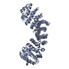
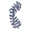
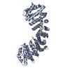
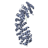
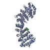
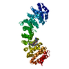
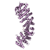
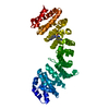
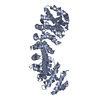
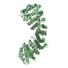
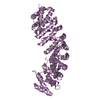
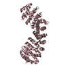
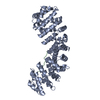
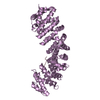
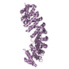
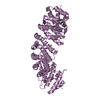
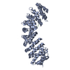
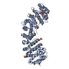
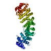
| Entry | Database: PDB / ID: 5svz | ||||||
|---|---|---|---|---|---|---|---|
| Title | HIV-1 Tat NLS in complex with importin alpha | ||||||
 Components Components |
| ||||||
 Keywords Keywords | Transport Protein/Viral Protein /  HIV-1 / Tat / Importin alpha / Virus / Complex / Transport Protein-Viral Protein complex HIV-1 / Tat / Importin alpha / Virus / Complex / Transport Protein-Viral Protein complex | ||||||
| Function / homology |  Function and homology information Function and homology information: / trans-activation response element binding / positive regulation of viral transcription / modulation by virus of host chromatin organization / Sensing of DNA Double Strand Breaks / entry of viral genome into host nucleus through nuclear pore complex via importin / positive regulation of viral life cycle / host cell nucleolus / NLS-dependent protein nuclear import complex / postsynapse to nucleus signaling pathway ...: / trans-activation response element binding / positive regulation of viral transcription / modulation by virus of host chromatin organization / Sensing of DNA Double Strand Breaks / entry of viral genome into host nucleus through nuclear pore complex via importin / positive regulation of viral life cycle / host cell nucleolus / NLS-dependent protein nuclear import complex / postsynapse to nucleus signaling pathway / actinin binding / nuclear import signal receptor activity / nuclear localization sequence binding / NLS-bearing protein import into nucleus / negative regulation of peptidyl-threonine phosphorylation / host cell / RNA-binding transcription regulator activity / cyclin binding / positive regulation of transcription elongation by RNA polymerase II / cytoplasmic stress granule / protein import into nucleus / histone deacetylase binding / nuclear membrane / DNA-binding transcription factor binding / host cell cytoplasm / postsynaptic density / protein domain specific binding / glutamatergic synapse / DNA-templated transcription / symbiont-mediated suppression of host type I interferon-mediated signaling pathway / extracellular region / nucleoplasm / metal ion binding / nucleus / cytosolSimilarity search - Function | ||||||
| Biological species |  Mus musculus (house mouse) Mus musculus (house mouse)  Human immunodeficiency virus 1 Human immunodeficiency virus 1 | ||||||
| Method | X-RAY DIFFRACTION / SYNCHROTRON / MOLECULAR REPLACEMENT / Resolution: 2 Å | ||||||
 Authors Authors | Smith, K.M. / Himiari, Z. / Forwood, J.K. | ||||||
 Citation Citation | Journal: Sci Rep / Year: 2017 Title: Structural Basis for Importin-alpha Binding of the Human Immunodeficiency Virus Tat. Authors: Smith, K.M. / Himiari, Z. / Tsimbalyuk, S. / Forwood, J.K. | ||||||
| History |
|
- Structure visualization
| Structure viewer | Molecule: MolmilJmol/JSmol |
|---|
- Downloads & links
-Download
| PDBx/mmCIF format | 5svz.cif.gz | 177.6 KB | Display | PDBx/mmCIF format |
|---|---|---|---|---|
| PDB format | pdb5svz.ent.gz | 140.4 KB | Display | PDB format |
| PDBx/mmJSON format | 5svz.json.gz | Tree view | PDBx/mmJSON format | |
| Others |  Other downloads Other downloads |
-Validation report
| Arichive directory | https://data.pdbj.org/pub/pdb/validation_reports/sv/5svzftp://data.pdbj.org/pub/pdb/validation_reports/sv/5svz | HTTPS FTP |
|---|
-Related structure data
| Related structure data |  5fc8S S: Starting model for refinement |
|---|---|
| Similar structure data |

-Links
 PDBj
PDBj


- Assembly
| Deposited unit | 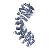
| ||||||||
|---|---|---|---|---|---|---|---|---|---|
| 1 |
| ||||||||
| Unit cell |
|
-Components
| #1: Protein | / Importin alpha P1 / Karyopherin subunit alpha-2 / Pendulin / Pore targeting complex 58 kDa subunit ...Importin alpha P1 / Karyopherin subunit alpha-2 / Pendulin / Pore targeting complex 58 kDa subunit / PTAC58 / RAG cohort protein 1 / SRP1-alpha Mass: 55268.473 Da / Num. of mol.: 1 Source method: isolated from a genetically manipulated source Source: (gene. exp.) Mus musculus (house mouse) / Gene: Kpna2, Rch1 / Production host:  Escherichia coli BL21(DE3) (bacteria) / References: UniProt: P52293 Escherichia coli BL21(DE3) (bacteria) / References: UniProt: P52293 |
|---|---|
| #2: Protein/peptide | Mass: 1902.220 Da / Num. of mol.: 1 / Fragment: NLS motif Source method: isolated from a genetically manipulated source Source: (gene. exp.) Human immunodeficiency virus 1 / Production host: Escherichia coli BL21(DE3) (bacteria) / References: UniProt: P04326*PLUS |
| #3: Water | ChemComp-HOH / Water Mass: 18.015 Da / Num. of mol.: 314 / Source method: isolated from a natural source / Formula: H2O Mass: 18.015 Da / Num. of mol.: 314 / Source method: isolated from a natural source / Formula: H2O |
-Experimental details
-Experiment
| Experiment | Method: X-RAY DIFFRACTION / Number of used crystals: 1 |
|---|
- Sample preparation
| Crystal | Density Matthews: 3.09 Å3/Da / Density % sol: 60.15 % |
|---|---|
| Crystal grow | Temperature: 296 K / Method: vapor diffusion, hanging drop / Details: 1.25 M sodium citrate pH 7 and 10 mM DTT |
-Data collection
| Diffraction | Mean temperature: 100 K | ||||||||||||||||||
|---|---|---|---|---|---|---|---|---|---|---|---|---|---|---|---|---|---|---|---|
| Diffraction source | Source: SYNCHROTRON / Site: Australian Synchrotron  / Beamline: MX2 / Wavelength: 0.9537 Å / Beamline: MX2 / Wavelength: 0.9537 Å | ||||||||||||||||||
| Detector | Type: ADSC QUANTUM 315r / Detector: CCD / Date: Jul 10, 2015 | ||||||||||||||||||
| Radiation | Protocol: SINGLE WAVELENGTH / Monochromatic (M) / Laue (L): M / Scattering type: x-ray | ||||||||||||||||||
| Radiation wavelength | Wavelength: 0.9537 Å / Relative weight: 1 | ||||||||||||||||||
| Reflection | Resolution: 2→29.662 Å / Num. obs: 46564 / % possible obs: 96.5 % / Redundancy: 4.6 % / Biso Wilson estimate: 27.42 Å2 / CC1/2: 0.998 / Rmerge(I) obs: 0.095 / Net I/σ(I): 13.4 | ||||||||||||||||||
| Reflection shell |
|
- Processing
| Software |
| ||||||||||||||||||||||||||||||||||||||||||||||||||||||||||||||||||||||||||||||||||||||||||||||||||||||||||||||||||||||||||||||
|---|---|---|---|---|---|---|---|---|---|---|---|---|---|---|---|---|---|---|---|---|---|---|---|---|---|---|---|---|---|---|---|---|---|---|---|---|---|---|---|---|---|---|---|---|---|---|---|---|---|---|---|---|---|---|---|---|---|---|---|---|---|---|---|---|---|---|---|---|---|---|---|---|---|---|---|---|---|---|---|---|---|---|---|---|---|---|---|---|---|---|---|---|---|---|---|---|---|---|---|---|---|---|---|---|---|---|---|---|---|---|---|---|---|---|---|---|---|---|---|---|---|---|---|---|---|---|---|
| Refinement | Method to determine structure: MOLECULAR REPLACEMENT Starting model: 5FC8 Resolution: 2→29.662 Å / SU ML: 0.19 / Cross valid method: FREE R-VALUE / σ(F): 1.34 / Phase error: 19.37
| ||||||||||||||||||||||||||||||||||||||||||||||||||||||||||||||||||||||||||||||||||||||||||||||||||||||||||||||||||||||||||||||
| Solvent computation | Shrinkage radii: 0.9 Å / VDW probe radii: 1.11 Å | ||||||||||||||||||||||||||||||||||||||||||||||||||||||||||||||||||||||||||||||||||||||||||||||||||||||||||||||||||||||||||||||
| Displacement parameters | Biso max: 136.04 Å2 / Biso mean: 40.434 Å2 / Biso min: 16.39 Å2 | ||||||||||||||||||||||||||||||||||||||||||||||||||||||||||||||||||||||||||||||||||||||||||||||||||||||||||||||||||||||||||||||
| Refinement step | Cycle: final / Resolution: 2→29.662 Å
| ||||||||||||||||||||||||||||||||||||||||||||||||||||||||||||||||||||||||||||||||||||||||||||||||||||||||||||||||||||||||||||||
| Refine LS restraints |
| ||||||||||||||||||||||||||||||||||||||||||||||||||||||||||||||||||||||||||||||||||||||||||||||||||||||||||||||||||||||||||||||
| LS refinement shell | Refine-ID: X-RAY DIFFRACTION / Total num. of bins used: 17
|
