 Movie
Movie Controller

[English] 日本語
 Yorodumi
- PDB-1elj: THE CRYSTAL STRUCTURE OF LIGANDED MALTODEXTRIN-BINDING PROTEIN FR... -
+ Open data

- Basic information
| Entry | Database: PDB / ID: 1elj | |||||||||
|---|---|---|---|---|---|---|---|---|---|---|
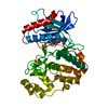
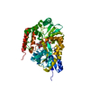
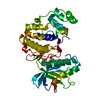
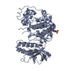
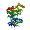
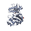
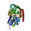
| Title | THE CRYSTAL STRUCTURE OF LIGANDED MALTODEXTRIN-BINDING PROTEIN FROM PYROCOCCUS FURIOSUS | |||||||||
 Components Components | MALTODEXTRIN-BINDING PROTEIN | |||||||||
 Keywords Keywords | SUGAR BINDING PROTEIN / protein-carbohydrate complex /  maltose binding protein / MBP fold / ABC transporter fold / thermophilic protein maltose binding protein / MBP fold / ABC transporter fold / thermophilic protein | |||||||||
| Function / homology |  Function and homology information Function and homology information | |||||||||
| Biological species |   Pyrococcus furiosus (archaea) Pyrococcus furiosus (archaea) | |||||||||
| Method | X-RAY DIFFRACTION / Resolution: 1.85 Å | |||||||||
 Authors Authors | Evdokimov, A.G. / Anderson, E.D. / Routzahn, K.M. / Waugh, D.S. | |||||||||
 Citation Citation | Journal: J.Mol.Biol. / Year: 2001 Title: Structural basis for oligosaccharide recognition by Pyrococcus furiosus maltodextrin-binding protein. Authors: Evdokimov, A.G. / Anderson, D.E. / Routzahn, K.M. / Waugh, D.S. | |||||||||
| History |
|
- Structure visualization
| Structure viewer | Molecule: MolmilJmol/JSmol |
|---|
- Downloads & links
-Download
| PDBx/mmCIF format | 1elj.cif.gz | 94.9 KB | Display | PDBx/mmCIF format |
|---|---|---|---|---|
| PDB format | pdb1elj.ent.gz | 74.1 KB | Display | PDB format |
| PDBx/mmJSON format | 1elj.json.gz | Tree view | PDBx/mmJSON format | |
| Others |  Other downloads Other downloads |
-Validation report
| Arichive directory | https://data.pdbj.org/pub/pdb/validation_reports/el/1eljftp://data.pdbj.org/pub/pdb/validation_reports/el/1elj | HTTPS FTP |
|---|
-Related structure data
| Related structure data | |
|---|---|
| Similar structure data |


-Links
 PDBj
PDBj


- Assembly
| Deposited unit | 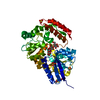
| ||||||||
|---|---|---|---|---|---|---|---|---|---|
| 1 |
| ||||||||
| Unit cell |
|
-Components
| #1: Protein | Mass: 43147.137 Da / Num. of mol.: 1 Source method: isolated from a genetically manipulated source Source: (gene. exp.) Pyrococcus furiosus (archaea) / Plasmid: PKM800 / Production host:  Escherichia coli (E. coli) / References: UniProt: P58300 Escherichia coli (E. coli) / References: UniProt: P58300 | ||||
|---|---|---|---|---|---|
| #2: Polysaccharide | alpha-D-glucopyranose-(1-4)-alpha-D-glucopyranose-(1-4)-alpha-D-glucopyranose / alpha-maltotriose  , Oligosaccharide / Class: Nutrient / Mass: 504.438 Da / Num. of mol.: 1 , Oligosaccharide / Class: Nutrient / Mass: 504.438 Da / Num. of mol.: 1Source method: isolated from a genetically manipulated source Details: oligosaccharide / References: alpha-maltotriose | ||||
| #3: Chemical | Sulfate  Mass: 96.063 Da / Num. of mol.: 3 / Source method: obtained synthetically / Formula: SO4 Mass: 96.063 Da / Num. of mol.: 3 / Source method: obtained synthetically / Formula: SO4#4: Water | ChemComp-HOH / | Water Mass: 18.015 Da / Num. of mol.: 387 / Source method: isolated from a natural source / Formula: H2O Mass: 18.015 Da / Num. of mol.: 387 / Source method: isolated from a natural source / Formula: H2ONonpolymer details | MALTOTRIOSE IS A TRISACCHARIDE, COMPOSED OF THREE GLUCOPYRANOSIDE UNITS LINKED BY ALPHA 1->4 BONDS. ...MALTOTRIOS | |
-Experimental details
-Experiment
| Experiment | Method: X-RAY DIFFRACTION / Number of used crystals: 1 |
|---|
- Sample preparation
| Crystal | Density Matthews: 3.11 Å3/Da / Density % sol: 60.47 % | |||||||||||||||||||||||||
|---|---|---|---|---|---|---|---|---|---|---|---|---|---|---|---|---|---|---|---|---|---|---|---|---|---|---|
| Crystal grow | Temperature: 298 K / Method: vapor diffusion, hanging drop / pH: 9 Details: reservoir: 2.4 M ammonium sulfate, 0.1 M bicine-hcl. drop: 6 mg/ml protein 1:1 with reservoir, pH 9.0, VAPOR DIFFUSION, HANGING DROP, temperature 298K | |||||||||||||||||||||||||
| Crystal grow | *PLUS | |||||||||||||||||||||||||
| Components of the solutions | *PLUS
|
-Data collection
| Diffraction | Mean temperature: 100 K |
|---|---|
| Diffraction source | Source: ROTATING ANODE / Type: RIGAKU / Wavelength: 1.54178 |
| Detector | Type: MARRESEARCH / Detector: IMAGE PLATE / Date: Oct 15, 1999 |
| Radiation | Protocol: SINGLE WAVELENGTH / Monochromatic (M) / Laue (L): M / Scattering type: x-ray |
| Radiation wavelength | Wavelength: 1.54178 Å / Relative weight: 1 |
| Reflection | Resolution: 1.85→100 Å / Num. all: 42983 / Num. obs: 35220 / % possible obs: 80 % / Observed criterion σ(F): 4 / Observed criterion σ(I): 2 / Redundancy: 4.5 % / Biso Wilson estimate: 25.3 Å2 / Rmerge(I) obs: 0.052 / Net I/σ(I): 20.08 |
| Reflection shell | Resolution: 1.85→1.9 Å / Redundancy: 3 % / Rmerge(I) obs: 0.23 / Num. unique all: 2996 / % possible all: 84.9 |
| Reflection | *PLUS Num. obs: 42983 / % possible obs: 91.88 % / Rmerge(I) obs: 0.053 |
- Processing
| Software |
| |||||||||||||||||||||||||
|---|---|---|---|---|---|---|---|---|---|---|---|---|---|---|---|---|---|---|---|---|---|---|---|---|---|---|
| Refinement | Resolution: 1.85→100 Å / σ(F): 4 / σ(I): 2 / Stereochemistry target values: Engh & Huber / Details: Used conjugated gradient least squares proce
| |||||||||||||||||||||||||
| Refinement step | Cycle: LAST / Resolution: 1.85→100 Å
| |||||||||||||||||||||||||
| Refine LS restraints |
| |||||||||||||||||||||||||
| Software | *PLUS Name: SHELXL-97 / Classification: refinement | |||||||||||||||||||||||||
| Refinement | *PLUS Lowest resolution: 100 Å / σ(F): 4 / % reflection Rfree: 5 % | |||||||||||||||||||||||||
| Solvent computation | *PLUS | |||||||||||||||||||||||||
| Displacement parameters | *PLUS | |||||||||||||||||||||||||
| Refine LS restraints | *PLUS
|
