 Movie
Movie Controller


+ Open data

- Basic information
| Entry | Database: PDB / ID: 3mbp | |||||||||
|---|---|---|---|---|---|---|---|---|---|---|
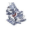
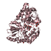
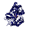
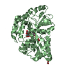
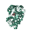
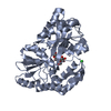
| Title | MALTODEXTRIN-BINDING PROTEIN WITH BOUND MALTOTRIOSE | |||||||||
 Components Components | MALTODEXTRIN-BINDING PROTEIN | |||||||||
 Keywords Keywords | PERIPLASMIC BINDING PROTEIN | |||||||||
| Function / homology |  Function and homology information Function and homology informationdetection of maltose stimulus /  maltose binding / maltose transport complex / maltose transport / maltodextrin transmembrane transport / carbohydrate transmembrane transporter activity / ATP-binding cassette (ABC) transporter complex, substrate-binding subunit-containing / carbohydrate transport / ATP-binding cassette (ABC) transporter complex / cell chemotaxis ...detection of maltose stimulus / maltose binding / maltose transport complex / maltose transport / maltodextrin transmembrane transport / carbohydrate transmembrane transporter activity / ATP-binding cassette (ABC) transporter complex, substrate-binding subunit-containing / carbohydrate transport / ATP-binding cassette (ABC) transporter complex / cell chemotaxis / outer membrane-bounded periplasmic space / periplasmic space / DNA damage response / membrane maltose binding / maltose transport complex / maltose transport / maltodextrin transmembrane transport / carbohydrate transmembrane transporter activity / ATP-binding cassette (ABC) transporter complex, substrate-binding subunit-containing / carbohydrate transport / ATP-binding cassette (ABC) transporter complex / cell chemotaxis ...detection of maltose stimulus / maltose binding / maltose transport complex / maltose transport / maltodextrin transmembrane transport / carbohydrate transmembrane transporter activity / ATP-binding cassette (ABC) transporter complex, substrate-binding subunit-containing / carbohydrate transport / ATP-binding cassette (ABC) transporter complex / cell chemotaxis / outer membrane-bounded periplasmic space / periplasmic space / DNA damage response / membraneSimilarity search - Function | |||||||||
| Biological species |  Escherichia coli (E. coli) Escherichia coli (E. coli) | |||||||||
| Method | X-RAY DIFFRACTION / ISOMORPHOUS WITH 2MBP / Resolution: 1.7 Å | |||||||||
 Authors Authors | Spurlino, J.C. / Quiocho, F.A. | |||||||||
 Citation Citation | Journal: Structure / Year: 1997 Title: Extensive features of tight oligosaccharide binding revealed in high-resolution structures of the maltodextrin transport/chemosensory receptor. Authors: Quiocho, F.A. / Spurlino, J.C. / Rodseth, L.E. #1: Journal: Biochemistry / Year: 1993Title: Refined 1.8-A Structure Reveals the Mode of Binding of Beta-Cyclodextrin to the Maltodextrin Binding Protein Authors: Sharff, A.J. / Rodseth, L.E. / Quiocho, F.A. #2: Journal: J.Mol.Biol. / Year: 1992Title: Atomic Interactions in Protein-Carbohydrate Complexes. Tryptophan Residues in the Periplasmic Maltodextrin Receptor for Active Transport and Chemotaxis Authors: Spurlino, J.C. / Rodseth, L.E. / Quiocho, F.A. #3: Journal: Biochemistry / Year: 1992Title: Crystallographic Evidence of a Large Ligand-Induced Hinge-Twist Motion between the Two Domains of the Maltodextrin Binding Protein Involved in Active Transport and Chemotaxis Authors: Sharff, A.J. / Rodseth, L.E. / Spurlino, J.C. / Quiocho, F.A. #4: Journal: J.Biol.Chem. / Year: 1991Title: The 2.3-A Resolution Structure of the Maltose-or Maltodextrin-Binding Protein, a Primary Receptor of Bacterial Active Transport and Chemotaxis Authors: Spurlino, J.C. / Lu, G.Y. / Quiocho, F.A. | |||||||||
| History |
|
- Structure visualization
| Structure viewer | Molecule: MolmilJmol/JSmol |
|---|
- Downloads & links
-Download
| PDBx/mmCIF format | 3mbp.cif.gz | 89.4 KB | Display | PDBx/mmCIF format |
|---|---|---|---|---|
| PDB format | pdb3mbp.ent.gz | 69.1 KB | Display | PDB format |
| PDBx/mmJSON format | 3mbp.json.gz | Tree view | PDBx/mmJSON format | |
| Others |  Other downloads Other downloads |
-Validation report
| Arichive directory | https://data.pdbj.org/pub/pdb/validation_reports/mb/3mbpftp://data.pdbj.org/pub/pdb/validation_reports/mb/3mbp | HTTPS FTP |
|---|
-Related structure data
| Related structure data |  1anfC  4mbpC  2mbp S: Starting model for refinement C: citing same article ( |
|---|---|
| Similar structure data |


-Links
 PDBj
PDBj


- Assembly
| Deposited unit | 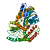
| ||||||||
|---|---|---|---|---|---|---|---|---|---|
| 1 |
| ||||||||
| Unit cell |
|
-Components
| #1: Protein | Mass: 40753.152 Da / Num. of mol.: 1 Source method: isolated from a genetically manipulated source Source: (gene. exp.) Escherichia coli (E. coli) / Strain: K12 / Cellular location: PERIPLASM / Gene: MALE / References: UniProt: P02928, UniProt: P0AEX9*PLUS |
|---|---|
| #2: Polysaccharide | alpha-D-glucopyranose-(1-4)-alpha-D-glucopyranose-(1-4)-alpha-D-glucopyranose / alpha-maltotriose  , Oligosaccharide / Class: Nutrient / Mass: 504.438 Da / Num. of mol.: 1 , Oligosaccharide / Class: Nutrient / Mass: 504.438 Da / Num. of mol.: 1Source method: isolated from a genetically manipulated source Details: oligosaccharide / References: alpha-maltotriose |
| #3: Water | ChemComp-HOH / Water Mass: 18.015 Da / Num. of mol.: 244 / Source method: isolated from a natural source / Formula: H2O Mass: 18.015 Da / Num. of mol.: 244 / Source method: isolated from a natural source / Formula: H2O |
-Experimental details
-Experiment
| Experiment | Method: X-RAY DIFFRACTION / Number of used crystals: 1 |
|---|
- Sample preparation
| Crystal | Density Matthews: 2.77 Å3/Da / Density % sol: 49 % | |||||||||||||||||||||||||
|---|---|---|---|---|---|---|---|---|---|---|---|---|---|---|---|---|---|---|---|---|---|---|---|---|---|---|
| Crystal grow | pH: 6.2 Details: PROTEIN WAS CRYSTALLIZED FROM 18% PEG 8000, 10 MM CITRATE, PH 6.2 | |||||||||||||||||||||||||
| Crystal grow | *PLUS Temperature: 4 ℃ / Method: vapor diffusion, hanging drop | |||||||||||||||||||||||||
| Components of the solutions | *PLUS
|
-Data collection
| Diffraction | Mean temperature: 287 K |
|---|---|
| Diffraction source | Source: ROTATING ANODE / Type: RIGAKU RUH2R / Wavelength: 1.5418 |
| Detector | Type: XUONG-HAMLIN MULTIWIRE / Detector: AREA DETECTOR / Date: Jan 1, 1989 / Details: NA |
| Radiation | Monochromator: GRAPHITE(002) / Monochromatic (M) / Laue (L): M / Scattering type: x-ray |
| Radiation wavelength | Wavelength: 1.5418 Å / Relative weight: 1 |
| Reflection | Resolution: 1.7→10 Å / Num. obs: 39297 / % possible obs: 86 % / Observed criterion σ(I): 1 / Redundancy: 3.7 % / Biso Wilson estimate: 19.5 Å2 / Rmerge(I) obs: 0.065 / Rsym value: 0.0969 / Net I/σ(I): 13.88 |
| Reflection shell | Resolution: 1.7→1.82 Å / Redundancy: 2.4 % / Rmerge(I) obs: 0.18 / Mean I/σ(I) obs: 2.5 / Rsym value: 0.244 / % possible all: 46 |
| Reflection shell | *PLUS % possible obs: 46 % |
- Processing
| Software |
| ||||||||||||||||||||||||||||||||||||||||||||||||||||||||||||||||||||||||||||||||||||
|---|---|---|---|---|---|---|---|---|---|---|---|---|---|---|---|---|---|---|---|---|---|---|---|---|---|---|---|---|---|---|---|---|---|---|---|---|---|---|---|---|---|---|---|---|---|---|---|---|---|---|---|---|---|---|---|---|---|---|---|---|---|---|---|---|---|---|---|---|---|---|---|---|---|---|---|---|---|---|---|---|---|---|---|---|---|
| Refinement | Method to determine structure: ISOMORPHOUS WITH 2MBP Starting model: PDB ENTRY 2MBP 2mbp Resolution: 1.7→10 Å / σ(F): 2 / Details: X-PLOR ALSO WAS USED.
| ||||||||||||||||||||||||||||||||||||||||||||||||||||||||||||||||||||||||||||||||||||
| Displacement parameters | Biso mean: 27.8 Å2 | ||||||||||||||||||||||||||||||||||||||||||||||||||||||||||||||||||||||||||||||||||||
| Refine analyze | Luzzati coordinate error obs: 0.2 Å / Luzzati d res low obs: 5 Å / Luzzati sigma a obs: 0.17 Å | ||||||||||||||||||||||||||||||||||||||||||||||||||||||||||||||||||||||||||||||||||||
| Refinement step | Cycle: LAST / Resolution: 1.7→10 Å
| ||||||||||||||||||||||||||||||||||||||||||||||||||||||||||||||||||||||||||||||||||||
| Refine LS restraints |
| ||||||||||||||||||||||||||||||||||||||||||||||||||||||||||||||||||||||||||||||||||||
| Software | *PLUS Name: PROLSQ / Classification: refinement | ||||||||||||||||||||||||||||||||||||||||||||||||||||||||||||||||||||||||||||||||||||
| Refinement | *PLUS Rfactor obs: 0.164 | ||||||||||||||||||||||||||||||||||||||||||||||||||||||||||||||||||||||||||||||||||||
| Solvent computation | *PLUS | ||||||||||||||||||||||||||||||||||||||||||||||||||||||||||||||||||||||||||||||||||||
| Displacement parameters | *PLUS |
