 Movie
Movie Controller

[English] 日本語
 Yorodumi
- PDB-4l2d: X-ray structure of the Fe(II) form of the iron superoxide dismuta... -
+ Open data

- Basic information
| Entry | Database: PDB / ID: 4l2d | |||||||||
|---|---|---|---|---|---|---|---|---|---|---|
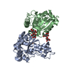
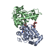
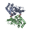
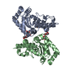
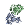
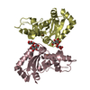
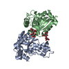
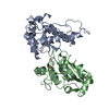
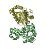
| Title | X-ray structure of the Fe(II) form of the iron superoxide dismutase from Pseudoalteromonas haloplanktis | |||||||||
 Components Components | Superoxide dismutase [Fe] | |||||||||
 Keywords Keywords |  OXIDOREDUCTASE / Superoxide dismutase OXIDOREDUCTASE / Superoxide dismutase | |||||||||
| Function / homology |  Function and homology informationsuperoxide dismutase / superoxide dismutase activity / metal ion binding Function and homology informationsuperoxide dismutase / superoxide dismutase activity / metal ion bindingSimilarity search - Function | |||||||||
| Biological species |  Pseudoalteromonas haloplanktis (bacteria) Pseudoalteromonas haloplanktis (bacteria) | |||||||||
| Method | X-RAY DIFFRACTION / MOLECULAR REPLACEMENT / Resolution: 2.07 Å | |||||||||
 Authors Authors | Russo Krauss, I. / Merlino, A. / Sica, F. | |||||||||
 Citation Citation | Journal: Biochim.Biophys.Acta / Year: 2014 Title: Structural and denaturation studies of two mutants of a cold adapted superoxide dismutase point to the importance of electrostatic interactions in protein stability. Authors: Merlino, A. / Russo Krauss, I. / Castellano, I. / Ruocco, M.R. / Capasso, A. / De Vendittis, E. / Rossi, B. / Sica, F. | |||||||||
| History |
|
- Structure visualization
| Structure viewer | Molecule: MolmilJmol/JSmol |
|---|
- Downloads & links
-Download
| PDBx/mmCIF format | 4l2d.cif.gz | 173.3 KB | Display | PDBx/mmCIF format |
|---|---|---|---|---|
| PDB format | pdb4l2d.ent.gz | 137.8 KB | Display | PDB format |
| PDBx/mmJSON format | 4l2d.json.gz | Tree view | PDBx/mmJSON format | |
| Others |  Other downloads Other downloads |
-Validation report
| Arichive directory | https://data.pdbj.org/pub/pdb/validation_reports/l2/4l2dftp://data.pdbj.org/pub/pdb/validation_reports/l2/4l2d | HTTPS FTP |
|---|
-Related structure data


-Links
 PDBj
PDBj


- Assembly
| Deposited unit | 
| ||||||||
|---|---|---|---|---|---|---|---|---|---|
| 1 | 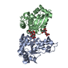
| ||||||||
| 2 | 
| ||||||||
| Unit cell |
|
-Components
| #1: Protein | Mass: 21269.508 Da / Num. of mol.: 4 Source method: isolated from a genetically manipulated source Source: (gene. exp.) Pseudoalteromonas haloplanktis (bacteria)Gene: sodB, PSHAa1215 / Production host: Escherichia coli (E. coli) / References: UniProt: P84612, superoxide dismutase#2: Polysaccharide | alpha-D-glucopyranose-(1-1)-alpha-D-glucopyranose / trehalose / 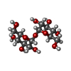  , Oligosaccharide / Class: Nutrient / Mass: 342.297 Da / Num. of mol.: 4 , Oligosaccharide / Class: Nutrient / Mass: 342.297 Da / Num. of mol.: 4Source method: isolated from a genetically manipulated source Details: oligosaccharide with reducing-end-to-reducing-end glycosidic bond References: trehalose#3: Chemical | ChemComp-FE2 /   Mass: 55.845 Da / Num. of mol.: 4 / Source method: obtained synthetically / Formula: Fe Mass: 55.845 Da / Num. of mol.: 4 / Source method: obtained synthetically / Formula: Fe#4: Water | ChemComp-HOH / | Water Mass: 18.015 Da / Num. of mol.: 623 / Source method: isolated from a natural source / Formula: H2O Mass: 18.015 Da / Num. of mol.: 623 / Source method: isolated from a natural source / Formula: H2O |
|---|
-Experimental details
-Experiment
| Experiment | Method: X-RAY DIFFRACTION / Number of used crystals: 1 |
|---|
- Sample preparation
| Crystal | Density Matthews: 2.66 Å3/Da / Density % sol: 53.75 % |
|---|---|
| Crystal grow | Temperature: 293 K / Method: vapor diffusion, hanging drop / pH: 7.5 Details: Ammonium sulphate 2.0 M, NaCl 0.1 M, Hepes 0.1 M, pH 7.5, VAPOR DIFFUSION, HANGING DROP, temperature 293K |
-Data collection
| Diffraction | Mean temperature: 100 K |
|---|---|
| Diffraction source | Source: ROTATING ANODE / Type: RIGAKU MICROMAX-007 HF / Wavelength: 1.54 Å |
| Radiation | Protocol: SINGLE WAVELENGTH / Monochromatic (M) / Laue (L): M / Scattering type: x-ray |
| Radiation wavelength | Wavelength: 1.54 Å / Relative weight: 1 |
| Reflection | Resolution: 2.07→29.2 Å / Num. all: 53308 / Num. obs: 53308 / % possible obs: 98.2 % / Observed criterion σ(F): 0 / Observed criterion σ(I): 0 |
| Reflection shell | Resolution: 2.07→2.14 Å / % possible all: 85.6 |
- Processing
| Software |
| ||||||||||||||||||
|---|---|---|---|---|---|---|---|---|---|---|---|---|---|---|---|---|---|---|---|
| Refinement | Method to determine structure: MOLECULAR REPLACEMENT / Resolution: 2.07→29.2 Å / σ(F): 0
| ||||||||||||||||||
| Refinement step | Cycle: LAST / Resolution: 2.07→29.2 Å
|
