 Movie
Movie Controller

[English] 日本語
 Yorodumi
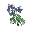
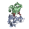
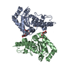
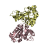
- PDB-3sdp: THE 2.1 ANGSTROMS RESOLUTION STRUCTURE OF IRON SUPEROXIDE DISMUTA... -
+ Open data

- Basic information
| Entry | Database: PDB / ID: 3sdp | ||||||
|---|---|---|---|---|---|---|---|
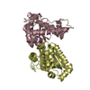
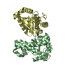
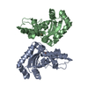
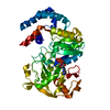
| Title | THE 2.1 ANGSTROMS RESOLUTION STRUCTURE OF IRON SUPEROXIDE DISMUTASE FROM PSEUDOMONAS OVALIS | ||||||
 Components Components | IRON SUPEROXIDE DISMUTASE | ||||||
 Keywords Keywords | OXIDOREDUCTASE (SUPEROXIDE ACCEPTOR) | ||||||
| Function / homology |  Function and homology informationsuperoxide dismutase / superoxide dismutase activity / metal ion binding Function and homology informationsuperoxide dismutase / superoxide dismutase activity / metal ion bindingSimilarity search - Function | ||||||
| Biological species |  Pseudomonas putida (bacteria) Pseudomonas putida (bacteria) | ||||||
| Method | X-RAY DIFFRACTION / Resolution: 2.1 Å | ||||||
 Authors Authors | Stoddard, B.L. / Ringe, D. / Petsko, G.A. | ||||||
 Citation Citation | Journal: Biochemistry / Year: 1990 Title: The 2.1-A resolution structure of iron superoxide dismutase from Pseudomonas ovalis. Authors: Stoddard, B.L. / Howell, P.L. / Ringe, D. / Petsko, G.A. #1: Journal: Protein Eng. / Year: 1990Title: The Structure of Iron Superoxide Dismutase from Pseudomonas Ovalis Bound to the Inhibitor Azide: Coordination Changes and Dynamics During Catalysis Authors: Stoddard, B.L. / Ringe, D. / Petsko, G.A. | ||||||
| History |
|
- Structure visualization
| Structure viewer | Molecule: MolmilJmol/JSmol |
|---|
- Downloads & links
-Download
| PDBx/mmCIF format | 3sdp.cif.gz | 85 KB | Display | PDBx/mmCIF format |
|---|---|---|---|---|
| PDB format | pdb3sdp.ent.gz | 60.3 KB | Display | PDB format |
| PDBx/mmJSON format | 3sdp.json.gz | Tree view | PDBx/mmJSON format | |
| Others |  Other downloads Other downloads |
-Validation report
| Arichive directory | https://data.pdbj.org/pub/pdb/validation_reports/sd/3sdpftp://data.pdbj.org/pub/pdb/validation_reports/sd/3sdp | HTTPS FTP |
|---|
-Related structure data
| Similar structure data |
|---|


-Links
 PDBj
PDBj
- Assembly
| Deposited unit | 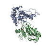
| ||||||||
|---|---|---|---|---|---|---|---|---|---|
| 1 |
| ||||||||
| Unit cell |
|
-Components
| #1: Protein | Mass: 21550.943 Da / Num. of mol.: 2 Source method: isolated from a genetically manipulated source Source: (gene. exp.) Pseudomonas putida (bacteria) / References: UniProt: P09223, superoxide dismutase#2: Chemical | Iron  Mass: 55.845 Da / Num. of mol.: 2 / Source method: obtained synthetically / Formula: Fe Mass: 55.845 Da / Num. of mol.: 2 / Source method: obtained synthetically / Formula: Fe#3: Water | ChemComp-HOH / | Water Mass: 18.015 Da / Num. of mol.: 180 / Source method: isolated from a natural source / Formula: H2O Mass: 18.015 Da / Num. of mol.: 180 / Source method: isolated from a natural source / Formula: H2OSequence details | RESIDUE 85 OF EACH CHAIN IS IDENTIFIED AS ALA IN THIS ENTRY. NOTE THAT THIS RESIDUE IS IDENTIFIED ...RESIDUE 85 OF EACH CHAIN IS IDENTIFIED | |
|---|
-Experimental details
-Experiment
| Experiment | Method: X-RAY DIFFRACTION |
|---|
- Sample preparation
| Crystal | Density Matthews: 2.75 Å3/Da / Density % sol: 55.34 % |
|---|---|
| Crystal grow | *PLUS Temperature: 4 ℃ / pH: 4.8 / Method: batch method |
| Components of the solutions | *PLUS Conc.: 55 %sat / Common name: ammonium sulfate |
-Data collection
| Reflection | *PLUS Highest resolution: 2.1 Å / Num. obs: 21140 / % possible obs: 72 % / Rmerge(I) obs: 0.086 |
|---|
- Processing
| Software |
| |||||||||||||||||||||||||||||||||||||||||||||||||||||||||||||||
|---|---|---|---|---|---|---|---|---|---|---|---|---|---|---|---|---|---|---|---|---|---|---|---|---|---|---|---|---|---|---|---|---|---|---|---|---|---|---|---|---|---|---|---|---|---|---|---|---|---|---|---|---|---|---|---|---|---|---|---|---|---|---|---|---|
| Refinement | Rfactor obs: 0.232 / Highest resolution: 2.1 Å | |||||||||||||||||||||||||||||||||||||||||||||||||||||||||||||||
| Refinement step | Cycle: LAST / Highest resolution: 2.1 Å
| |||||||||||||||||||||||||||||||||||||||||||||||||||||||||||||||
| Refine LS restraints |
| |||||||||||||||||||||||||||||||||||||||||||||||||||||||||||||||
| Software | *PLUS Name: PROLSQ / Classification: refinement | |||||||||||||||||||||||||||||||||||||||||||||||||||||||||||||||
| Refine LS restraints | *PLUS
|
