 Movie
Movie Controller

[English] 日本語
 Yorodumi
- PDB-4k7k: Crystal structures of CusC review conformational changes accompan... -
+ Open data

- Basic information
| Entry | Database: PDB / ID: 4k7k | ||||||
|---|---|---|---|---|---|---|---|
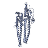
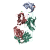
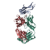
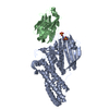
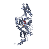
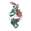
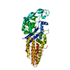
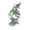
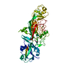
| Title | Crystal structures of CusC review conformational changes accompanying folding and transmembrane channel formation | ||||||
 Components Components | Cation efflux system protein CusC | ||||||
 Keywords Keywords |  MEMBRANE PROTEIN / beta barrel MEMBRANE PROTEIN / beta barrel | ||||||
| Function / homology |  Function and homology information Function and homology informationprotein palmitoylation / copper ion transmembrane transport / response to silver ion / silver ion transmembrane transport / diacylglycerol binding / copper ion transmembrane transporter activity / copper ion export / detoxification of copper ion / response to copper ion / porin activity ...protein palmitoylation / copper ion transmembrane transport / response to silver ion / silver ion transmembrane transport / diacylglycerol binding / copper ion transmembrane transporter activity / copper ion export / detoxification of copper ion / response to copper ion / porin activity / pore complex / efflux transmembrane transporter activity / protein homotrimerization / transmembrane transporter activity / intracellular copper ion homeostasis / cell outer membrane / transmembrane transport / response to toxic substance / copper ion binding / membrane / identical protein bindingSimilarity search - Function | ||||||
| Biological species |  Escherichia coli (E. coli) Escherichia coli (E. coli) | ||||||
| Method | X-RAY DIFFRACTION / SYNCHROTRON / SAD / Resolution: 2.53 Å | ||||||
 Authors Authors | Su, C.-C. / Lei, H.-T. | ||||||
 Citation Citation | Journal: J.Mol.Biol. / Year: 2014 Title: Crystal Structures of CusC Review Conformational Changes Accompanying Folding and Transmembrane Channel Formation. Authors: Lei, H.T. / Bolla, J.R. / Bishop, N.R. / Su, C.C. / Yu, E.W. | ||||||
| History |
|
- Structure visualization
| Structure viewer | Molecule: MolmilJmol/JSmol |
|---|
- Downloads & links
-Download
| PDBx/mmCIF format | 4k7k.cif.gz | 155.9 KB | Display | PDBx/mmCIF format |
|---|---|---|---|---|
| PDB format | pdb4k7k.ent.gz | 123.4 KB | Display | PDB format |
| PDBx/mmJSON format | 4k7k.json.gz | Tree view | PDBx/mmJSON format | |
| Others |  Other downloads Other downloads |
-Validation report
| Arichive directory | https://data.pdbj.org/pub/pdb/validation_reports/k7/4k7kftp://data.pdbj.org/pub/pdb/validation_reports/k7/4k7k | HTTPS FTP |
|---|
-Related structure data


-Links
 PDBj
PDBj


- Assembly
| Deposited unit | 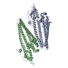
| ||||||||
|---|---|---|---|---|---|---|---|---|---|
| 1 | 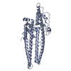
| ||||||||
| 2 | 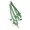
| ||||||||
| Unit cell |
|
-Components
| #1: Protein | Mass: 49391.977 Da / Num. of mol.: 2 / Fragment: UNP residues 18-457 Source method: isolated from a genetically manipulated source Source: (gene. exp.) Escherichia coli (E. coli) / Strain: K-12 / Gene: cusC, ibeB, ylcB, b0572, JW0561 / Plasmid: pBAD22 / Production host: Escherichia coli (E. coli) / References: UniProt: P77211#2: Water | ChemComp-HOH / | Water Mass: 18.015 Da / Num. of mol.: 88 / Source method: isolated from a natural source / Formula: H2O Mass: 18.015 Da / Num. of mol.: 88 / Source method: isolated from a natural source / Formula: H2O |
|---|
-Experimental details
-Experiment
| Experiment | Method: X-RAY DIFFRACTION / Number of used crystals: 1 |
|---|
- Sample preparation
| Crystal | Density Matthews: 2.32 Å3/Da / Density % sol: 46.97 % |
|---|---|
| Crystal grow | Temperature: 298 K / Method: vapor diffusion / pH: 7.5 Details: 7% PEG3350, 0.2M NH4SO4, 0.1M HEPES (7.5), vapor diffusion, temperature 298K |
-Data collection
| Diffraction | Mean temperature: 100 K | ||||||||||||||||||||||||||||||||||||||||||||||||||||||||||||||||||||||
|---|---|---|---|---|---|---|---|---|---|---|---|---|---|---|---|---|---|---|---|---|---|---|---|---|---|---|---|---|---|---|---|---|---|---|---|---|---|---|---|---|---|---|---|---|---|---|---|---|---|---|---|---|---|---|---|---|---|---|---|---|---|---|---|---|---|---|---|---|---|---|---|
| Diffraction source | Source: SYNCHROTRON / Site: APS  / Beamline: 24-ID-E / Wavelength: 0.98 Å / Beamline: 24-ID-E / Wavelength: 0.98 Å | ||||||||||||||||||||||||||||||||||||||||||||||||||||||||||||||||||||||
| Detector | Type: ADSC QUANTUM 315 / Detector: CCD / Date: Feb 3, 2012 | ||||||||||||||||||||||||||||||||||||||||||||||||||||||||||||||||||||||
| Radiation | Monochromator: Si(III) / Protocol: SINGLE WAVELENGTH / Monochromatic (M) / Laue (L): M / Scattering type: x-ray | ||||||||||||||||||||||||||||||||||||||||||||||||||||||||||||||||||||||
| Radiation wavelength | Wavelength: 0.98 Å / Relative weight: 1 | ||||||||||||||||||||||||||||||||||||||||||||||||||||||||||||||||||||||
| Reflection | Resolution: 2.53→50 Å / Num. obs: 30128 / % possible obs: 94 % / Observed criterion σ(F): 2 / Observed criterion σ(I): 2 / Redundancy: 2 % / Rmerge(I) obs: 0.069 / Χ2: 0.994 / Net I/σ(I): 8 | ||||||||||||||||||||||||||||||||||||||||||||||||||||||||||||||||||||||
| Reflection shell |
|
- Processing
| Software |
| ||||||||||||||||||||||||||||||||||||||||||||||||||||||||||||||||||||||||||||||||||||
|---|---|---|---|---|---|---|---|---|---|---|---|---|---|---|---|---|---|---|---|---|---|---|---|---|---|---|---|---|---|---|---|---|---|---|---|---|---|---|---|---|---|---|---|---|---|---|---|---|---|---|---|---|---|---|---|---|---|---|---|---|---|---|---|---|---|---|---|---|---|---|---|---|---|---|---|---|---|---|---|---|---|---|---|---|---|
| Refinement | Method to determine structure: SAD / Resolution: 2.53→46.011 Å / Occupancy max: 1 / Occupancy min: 1 / FOM work R set: 0.7956 / SU ML: 0.4 / σ(F): 1.34 / Phase error: 27.71 / Stereochemistry target values: ML
| ||||||||||||||||||||||||||||||||||||||||||||||||||||||||||||||||||||||||||||||||||||
| Solvent computation | Shrinkage radii: 0.98 Å / VDW probe radii: 1.2 Å / Solvent model: FLAT BULK SOLVENT MODEL / Bsol: 53.776 Å2 / ksol: 0.375 e/Å3 | ||||||||||||||||||||||||||||||||||||||||||||||||||||||||||||||||||||||||||||||||||||
| Displacement parameters | Biso max: 132.37 Å2 / Biso mean: 47.8236 Å2 / Biso min: 15.87 Å2
| ||||||||||||||||||||||||||||||||||||||||||||||||||||||||||||||||||||||||||||||||||||
| Refinement step | Cycle: LAST / Resolution: 2.53→46.011 Å
| ||||||||||||||||||||||||||||||||||||||||||||||||||||||||||||||||||||||||||||||||||||
| Refine LS restraints |
| ||||||||||||||||||||||||||||||||||||||||||||||||||||||||||||||||||||||||||||||||||||
| LS refinement shell | Refine-ID: X-RAY DIFFRACTION / Total num. of bins used: 11
|
