 Movie
Movie Controller

[English] 日本語
 Yorodumi
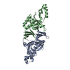
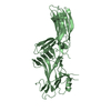
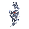
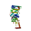
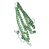
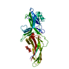
- PDB-5nf3: The fimbrial shaft protein Mfa1 from Porphyromonas gingivalis-C-t... -
+ Open data

- Basic information
| Entry | Database: PDB / ID: 5nf3 | |||||||||
|---|---|---|---|---|---|---|---|---|---|---|
| Title | The fimbrial shaft protein Mfa1 from Porphyromonas gingivalis-C-terminal deletion | |||||||||
 Components Components | Minor fimbrium subunit Mfa1 | |||||||||
 Keywords Keywords |  CELL ADHESION / FIMBRIA / ADHESIN / PERIODONTITIS CELL ADHESION / FIMBRIA / ADHESIN / PERIODONTITIS | |||||||||
| Function / homology | pilus shaft / Fimbrial subunit protein, C-terminal / Major fimbrial subunit protein type IV, Fimbrillin, C-terminal / outer membrane / cell outer membrane / cell-cell adhesion / Prokaryotic membrane lipoprotein lipid attachment site profile. / ACETATE ION / Minor fimbrium subunit Mfa1 Function and homology information Function and homology information | |||||||||
| Biological species |  Porphyromonas gingivalis ATCC 33277 (bacteria) Porphyromonas gingivalis ATCC 33277 (bacteria) | |||||||||
| Method | X-RAY DIFFRACTION / SYNCHROTRON / MOLECULAR REPLACEMENT / Resolution: 1.97 Å | |||||||||
 Authors Authors | Hall, M. / Hasegawa, Y. / Persson, K. / Yoshimura, F. | |||||||||
| Funding support |  Sweden, Sweden,  Japan, 2items Japan, 2items
| |||||||||
 Citation Citation | Journal: Sci Rep / Year: 2018 Title: Structural and functional characterization of shaft, anchor, and tip proteins of the Mfa1 fimbria from the periodontal pathogen Porphyromonas gingivalis. Authors: Hall, M. / Hasegawa, Y. / Yoshimura, F. / Persson, K. | |||||||||
| History |
|
- Structure visualization
| Structure viewer | Molecule: MolmilJmol/JSmol |
|---|
- Downloads & links
-Download
| PDBx/mmCIF format | 5nf3.cif.gz | 289.4 KB | Display | PDBx/mmCIF format |
|---|---|---|---|---|
| PDB format | pdb5nf3.ent.gz | 234.8 KB | Display | PDB format |
| PDBx/mmJSON format | 5nf3.json.gz | Tree view | PDBx/mmJSON format | |
| Others |  Other downloads Other downloads |
-Validation report
| Arichive directory | https://data.pdbj.org/pub/pdb/validation_reports/nf/5nf3ftp://data.pdbj.org/pub/pdb/validation_reports/nf/5nf3 | HTTPS FTP |
|---|
-Related structure data


-Links
 PDBj
PDBj


- Assembly
| Deposited unit | 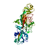
| ||||||||
|---|---|---|---|---|---|---|---|---|---|
| 1 |
| ||||||||
| Unit cell |
| ||||||||
| Components on special symmetry positions |
|
-Components
| #1: Protein | Mass: 56536.090 Da / Num. of mol.: 1 Source method: isolated from a genetically manipulated source Source: (gene. exp.) Porphyromonas gingivalis ATCC 33277 (bacteria)Gene: mfa1, PGN_0287 / Production host: Escherichia coli BL21 (bacteria) / References: UniProt: B2RHG1 |
|---|---|
| #2: Chemical | ChemComp-CA /   Mass: 40.078 Da / Num. of mol.: 1 / Source method: obtained synthetically / Formula: Ca Mass: 40.078 Da / Num. of mol.: 1 / Source method: obtained synthetically / Formula: Ca |
| #3: Chemical | ChemComp-ACT / Acetate  Mass: 59.044 Da / Num. of mol.: 1 / Source method: obtained synthetically / Formula: C2H3O2 Mass: 59.044 Da / Num. of mol.: 1 / Source method: obtained synthetically / Formula: C2H3O2 |
| #4: Water | ChemComp-HOH / Water Mass: 18.015 Da / Num. of mol.: 300 / Source method: isolated from a natural source / Formula: H2O Mass: 18.015 Da / Num. of mol.: 300 / Source method: isolated from a natural source / Formula: H2O |
-Experimental details
-Experiment
| Experiment | Method: X-RAY DIFFRACTION / Number of used crystals: 1 |
|---|
- Sample preparation
| Crystal | Density Matthews: 2.78 Å3/Da / Density % sol: 55.79 % |
|---|---|
| Crystal grow | Temperature: 293 K / Method: vapor diffusion, sitting drop / pH: 6.5 Details: 20% PEG 8000, 75 mM calcium acetate, 0.1 M sodium cacodylate pH 6.5. |
-Data collection
| Diffraction | Mean temperature: 100 K | |||||||||||||||||||||
|---|---|---|---|---|---|---|---|---|---|---|---|---|---|---|---|---|---|---|---|---|---|---|
| Diffraction source | Source: SYNCHROTRON / Site: ESRF  / Beamline: ID23-1 / Wavelength: 0.9794 Å / Beamline: ID23-1 / Wavelength: 0.9794 Å | |||||||||||||||||||||
| Detector | Type: DECTRIS PILATUS3 6M / Detector: PIXEL / Date: Mar 8, 2016 | |||||||||||||||||||||
| Radiation | Protocol: SINGLE WAVELENGTH / Monochromatic (M) / Laue (L): M / Scattering type: x-ray | |||||||||||||||||||||
| Radiation wavelength | Wavelength: 0.9794 Å / Relative weight: 1 | |||||||||||||||||||||
| Reflection | Resolution: 1.97→48.65 Å / Num. obs: 46649 / % possible obs: 100 % / Redundancy: 12.6 % / Biso Wilson estimate: 25.76 Å2 / CC1/2: 0.997 / Rmerge(I) obs: 0.211 / Rpim(I) all: 0.061 / Rrim(I) all: 0.22 / Net I/σ(I): 14.9 / Num. measured all: 587519 / Scaling rejects: 1543 | |||||||||||||||||||||
| Reflection shell | Diffraction-ID: 1
|
- Processing
| Software |
| ||||||||||||||||||||||||||||||||||||||||||||||||||||||||||||||||||||||||||||||||||||||||||||||||||||
|---|---|---|---|---|---|---|---|---|---|---|---|---|---|---|---|---|---|---|---|---|---|---|---|---|---|---|---|---|---|---|---|---|---|---|---|---|---|---|---|---|---|---|---|---|---|---|---|---|---|---|---|---|---|---|---|---|---|---|---|---|---|---|---|---|---|---|---|---|---|---|---|---|---|---|---|---|---|---|---|---|---|---|---|---|---|---|---|---|---|---|---|---|---|---|---|---|---|---|---|---|---|
| Refinement | Method to determine structure: MOLECULAR REPLACEMENT / Resolution: 1.97→48.648 Å / SU ML: 0.27 / Cross valid method: FREE R-VALUE / σ(F): 1.34 / Phase error: 19.51
| ||||||||||||||||||||||||||||||||||||||||||||||||||||||||||||||||||||||||||||||||||||||||||||||||||||
| Solvent computation | Shrinkage radii: 0.9 Å / VDW probe radii: 1.11 Å | ||||||||||||||||||||||||||||||||||||||||||||||||||||||||||||||||||||||||||||||||||||||||||||||||||||
| Displacement parameters | Biso max: 169.35 Å2 / Biso mean: 36.3687 Å2 / Biso min: 13.33 Å2 | ||||||||||||||||||||||||||||||||||||||||||||||||||||||||||||||||||||||||||||||||||||||||||||||||||||
| Refinement step | Cycle: final / Resolution: 1.97→48.648 Å
| ||||||||||||||||||||||||||||||||||||||||||||||||||||||||||||||||||||||||||||||||||||||||||||||||||||
| Refine LS restraints |
| ||||||||||||||||||||||||||||||||||||||||||||||||||||||||||||||||||||||||||||||||||||||||||||||||||||
| LS refinement shell | Refine-ID: X-RAY DIFFRACTION / Total num. of bins used: 14 / % reflection obs: 100 %
| ||||||||||||||||||||||||||||||||||||||||||||||||||||||||||||||||||||||||||||||||||||||||||||||||||||
| Refinement TLS params. | Method: refined / Refine-ID: X-RAY DIFFRACTION
| ||||||||||||||||||||||||||||||||||||||||||||||||||||||||||||||||||||||||||||||||||||||||||||||||||||
| Refinement TLS group |
|
