 Movie
Movie Controller

[English] 日本語
 Yorodumi
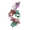
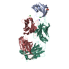
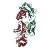
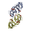
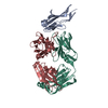
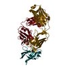
- PDB-6ddm: Crystal Structure Analysis of the Epitope of an Anti-MICA Antibody -
+ Open data

- Basic information
| Entry | Database: PDB / ID: 6ddm | ||||||
|---|---|---|---|---|---|---|---|
| Title | Crystal Structure Analysis of the Epitope of an Anti-MICA Antibody | ||||||
 Components Components |
| ||||||
 Keywords Keywords |  IMMUNE SYSTEM / Fab fragment-antigen complex / immunoglobulin domain IMMUNE SYSTEM / Fab fragment-antigen complex / immunoglobulin domain | ||||||
| Function / homology |  Function and homology information Function and homology informationantigen processing and presentation of endogenous peptide antigen via MHC class I via ER pathway, TAP-independent / antigen processing and presentation of endogenous peptide antigen via MHC class Ib / positive regulation of T cell mediated cytotoxicity / peptide antigen binding / immune response / external side of plasma membrane / signaling receptor binding / extracellular space / metal ion bindingSimilarity search - Function | ||||||
| Biological species |  Mus musculus (house mouse) Mus musculus (house mouse) Homo sapiens (human) Homo sapiens (human) | ||||||
| Method | X-RAY DIFFRACTION / SYNCHROTRON / MOLECULAR REPLACEMENT / Resolution: 1.3 Å | ||||||
 Authors Authors | Matsumoto, M.L. | ||||||
 Citation Citation | Journal: MAbs / Year: 2019 Title: High-resolution glycosylation site-engineering method identifies MICA epitope critical for shedding inhibition activity of anti-MICA antibodies. Authors: Lombana, T.N. / Matsumoto, M.L. / Berkley, A.M. / Toy, E. / Cook, R. / Gan, Y. / Du, C. / Schnier, P. / Sandoval, W. / Ye, Z. / Schartner, J.M. / Kim, J. / Spiess, C. | ||||||
| History |
|
- Structure visualization
| Structure viewer | Molecule: MolmilJmol/JSmol |
|---|
- Downloads & links
-Download
| PDBx/mmCIF format | 6ddm.cif.gz | 229.2 KB | Display | PDBx/mmCIF format |
|---|---|---|---|---|
| PDB format | pdb6ddm.ent.gz | 181.3 KB | Display | PDB format |
| PDBx/mmJSON format | 6ddm.json.gz | Tree view | PDBx/mmJSON format | |
| Others |  Other downloads Other downloads |
-Validation report
| Arichive directory | https://data.pdbj.org/pub/pdb/validation_reports/dd/6ddmftp://data.pdbj.org/pub/pdb/validation_reports/dd/6ddm | HTTPS FTP |
|---|
-Related structure data
| Related structure data |  6ddrC  6ddvC  1f3dS 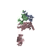 1hyrS  1nz8S 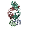 4m1gS S: Starting model for refinement C: citing same article ( |
|---|---|
| Similar structure data |


-Links
 PDBj
PDBj

- Assembly
| Deposited unit | 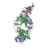
| ||||||||
|---|---|---|---|---|---|---|---|---|---|
| 1 |
| ||||||||
| Unit cell |
|
-Components
| #1: Antibody | Mass: 23219.854 Da / Num. of mol.: 1 Source method: isolated from a genetically manipulated source Details: chimeric Fab fragment consisting of murine variable domains with human constant domains Source: (gene. exp.) Mus musculus (house mouse) / Strain: Balb/c / Plasmid: pBR322 / Production host:  Escherichia coli (E. coli) / Strain (production host): 64B4 Escherichia coli (E. coli) / Strain (production host): 64B4 |
|---|---|
| #2: Protein | Mass: 10578.635 Da / Num. of mol.: 1 Source method: isolated from a genetically manipulated source Details: alpha 3 domain of the MICA*008 allele co-expressed with Streptomyces plicatu EndoH in the presence of kifunensine Source: (gene. exp.) Homo sapiens (human) / Gene: MICA, hCG_2001511 / Plasmid: pAcgp67 / Production host:  Trichoplusia ni (cabbage looper) / References: UniProt: Q96QC4 Trichoplusia ni (cabbage looper) / References: UniProt: Q96QC4 |
| #3: Antibody | Mass: 23441.264 Da / Num. of mol.: 1 Source method: isolated from a genetically manipulated source Details: himeric Fab fragment consisting of murine variable domains with human constant domains Source: (gene. exp.) Mus musculus (house mouse) / Plasmid: pBR322 / Production host: Escherichia coli (E. coli) |
| #4: Water | ChemComp-HOH / Water Mass: 18.015 Da / Num. of mol.: 618 / Source method: isolated from a natural source / Formula: H2O Mass: 18.015 Da / Num. of mol.: 618 / Source method: isolated from a natural source / Formula: H2O |
-Experimental details
-Experiment
| Experiment | Method: X-RAY DIFFRACTION / Number of used crystals: 1 |
|---|
- Sample preparation
| Crystal | Density Matthews: 2.28 Å3/Da / Density % sol: 46.17 % / Mosaicity: 0.498 ° |
|---|---|
| Crystal grow | Temperature: 277 K / Method: vapor diffusion, hanging drop / pH: 4.6 / Details: 0.1 M sodium acetate, pH 4.6, 25% PEG 4000 |
-Data collection
| Diffraction | Mean temperature: 100 K | |||||||||||||||||||||||||||||||||||||||||||||||||||||||||||||||||||||||||||||||||||||||||||||||||||||||||||||||||||||||||||||||||||||||||||||||||||||||||||||||||||||||||||||||||||||||||||||
|---|---|---|---|---|---|---|---|---|---|---|---|---|---|---|---|---|---|---|---|---|---|---|---|---|---|---|---|---|---|---|---|---|---|---|---|---|---|---|---|---|---|---|---|---|---|---|---|---|---|---|---|---|---|---|---|---|---|---|---|---|---|---|---|---|---|---|---|---|---|---|---|---|---|---|---|---|---|---|---|---|---|---|---|---|---|---|---|---|---|---|---|---|---|---|---|---|---|---|---|---|---|---|---|---|---|---|---|---|---|---|---|---|---|---|---|---|---|---|---|---|---|---|---|---|---|---|---|---|---|---|---|---|---|---|---|---|---|---|---|---|---|---|---|---|---|---|---|---|---|---|---|---|---|---|---|---|---|---|---|---|---|---|---|---|---|---|---|---|---|---|---|---|---|---|---|---|---|---|---|---|---|---|---|---|---|---|---|---|---|---|
| Diffraction source | Source: SYNCHROTRON / Site: APS  / Beamline: 22-ID / Wavelength: 1 Å / Beamline: 22-ID / Wavelength: 1 Å | |||||||||||||||||||||||||||||||||||||||||||||||||||||||||||||||||||||||||||||||||||||||||||||||||||||||||||||||||||||||||||||||||||||||||||||||||||||||||||||||||||||||||||||||||||||||||||||
| Detector | Type: RAYONIX MX300-HS / Detector: CCD / Date: Oct 14, 2015 | |||||||||||||||||||||||||||||||||||||||||||||||||||||||||||||||||||||||||||||||||||||||||||||||||||||||||||||||||||||||||||||||||||||||||||||||||||||||||||||||||||||||||||||||||||||||||||||
| Radiation | Protocol: SINGLE WAVELENGTH / Monochromatic (M) / Laue (L): M / Scattering type: x-ray | |||||||||||||||||||||||||||||||||||||||||||||||||||||||||||||||||||||||||||||||||||||||||||||||||||||||||||||||||||||||||||||||||||||||||||||||||||||||||||||||||||||||||||||||||||||||||||||
| Radiation wavelength | Wavelength: 1 Å / Relative weight: 1 | |||||||||||||||||||||||||||||||||||||||||||||||||||||||||||||||||||||||||||||||||||||||||||||||||||||||||||||||||||||||||||||||||||||||||||||||||||||||||||||||||||||||||||||||||||||||||||||
| Reflection | Resolution: 1.3→35 Å / Num. obs: 126363 / % possible obs: 99.6 % / Redundancy: 3.5 % / Rmerge(I) obs: 0.059 / Rpim(I) all: 0.037 / Rrim(I) all: 0.07 / Χ2: 0.997 / Net I/σ(I): 8.5 | |||||||||||||||||||||||||||||||||||||||||||||||||||||||||||||||||||||||||||||||||||||||||||||||||||||||||||||||||||||||||||||||||||||||||||||||||||||||||||||||||||||||||||||||||||||||||||||
| Reflection shell | Diffraction-ID: 1
|
- Processing
| Software |
| ||||||||||||||||||||||||||||||||||||||||||||||||||||||||||||||||||||||||||||||||||||||||||||||||||||
|---|---|---|---|---|---|---|---|---|---|---|---|---|---|---|---|---|---|---|---|---|---|---|---|---|---|---|---|---|---|---|---|---|---|---|---|---|---|---|---|---|---|---|---|---|---|---|---|---|---|---|---|---|---|---|---|---|---|---|---|---|---|---|---|---|---|---|---|---|---|---|---|---|---|---|---|---|---|---|---|---|---|---|---|---|---|---|---|---|---|---|---|---|---|---|---|---|---|---|---|---|---|
| Refinement | Method to determine structure: MOLECULAR REPLACEMENT Starting model: 1F3D, 4M1G, 1HYR, 1NZ8 Resolution: 1.3→35 Å / Cor.coef. Fo:Fc: 0.969 / Cor.coef. Fo:Fc free: 0.964 / SU B: 2.755 / SU ML: 0.054 / Cross valid method: THROUGHOUT / σ(F): 0 / ESU R: 0.054 / ESU R Free: 0.054 Details: U VALUES : WITH TLS ADDED HYDROGENS HAVE BEEN ADDED IN THE RIDING POSITIONS
| ||||||||||||||||||||||||||||||||||||||||||||||||||||||||||||||||||||||||||||||||||||||||||||||||||||
| Solvent computation | Ion probe radii: 0.8 Å / Shrinkage radii: 0.8 Å / VDW probe radii: 1.2 Å | ||||||||||||||||||||||||||||||||||||||||||||||||||||||||||||||||||||||||||||||||||||||||||||||||||||
| Displacement parameters | Biso max: 67.47 Å2 / Biso mean: 20.983 Å2 / Biso min: 11.7 Å2
| ||||||||||||||||||||||||||||||||||||||||||||||||||||||||||||||||||||||||||||||||||||||||||||||||||||
| Refinement step | Cycle: final / Resolution: 1.3→35 Å
| ||||||||||||||||||||||||||||||||||||||||||||||||||||||||||||||||||||||||||||||||||||||||||||||||||||
| Refine LS restraints |
| ||||||||||||||||||||||||||||||||||||||||||||||||||||||||||||||||||||||||||||||||||||||||||||||||||||
| LS refinement shell | Resolution: 1.3→1.334 Å / Rfactor Rfree error: 0 / Total num. of bins used: 20
| ||||||||||||||||||||||||||||||||||||||||||||||||||||||||||||||||||||||||||||||||||||||||||||||||||||
| Refinement TLS params. | Method: refined / Refine-ID: X-RAY DIFFRACTION
| ||||||||||||||||||||||||||||||||||||||||||||||||||||||||||||||||||||||||||||||||||||||||||||||||||||
| Refinement TLS group |
|
