 Movie
Movie Controller


+ Open data

- Basic information
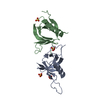
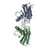
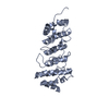
| Entry | Database: PDB / ID: 4k1o | ||||||
|---|---|---|---|---|---|---|---|
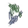
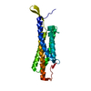
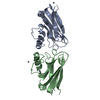
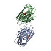
| Title | Crystal structure of the alphaN-catenin actin-binding domain | ||||||
 Components Components | Catenin alpha-2 CTNNA2 CTNNA2 | ||||||
 Keywords Keywords | CELL ADHESION / five-helix bundle / F-actin / alpha-catenin | ||||||
| Function / homology |  Function and homology information Function and homology informationradial glia guided migration of Purkinje cell / extrinsic component of postsynaptic membrane / regulation of synapse structural plasticity / extrinsic component of presynaptic membrane / modification of postsynaptic actin cytoskeleton / negative regulation of Arp2/3 complex-mediated actin nucleation / regulation of neuron migration / presynaptic active zone cytoplasmic component / Myogenesis / brain morphogenesis ...radial glia guided migration of Purkinje cell / extrinsic component of postsynaptic membrane / regulation of synapse structural plasticity / extrinsic component of presynaptic membrane / modification of postsynaptic actin cytoskeleton / negative regulation of Arp2/3 complex-mediated actin nucleation / regulation of neuron migration / presynaptic active zone cytoplasmic component / Myogenesis / brain morphogenesis / catenin complex / parallel fiber to Purkinje cell synapse / dendrite morphogenesis / regulation of neuron projection development / postsynaptic density, intracellular component / prepulse inhibition / hippocampal mossy fiber to CA3 synapse / axonogenesis / adherens junction / cell-cell adhesion / beta-catenin binding / cell migration / actin filament binding / actin cytoskeleton / lamellipodium / basolateral plasma membrane / postsynaptic density / cadherin binding / axon / structural molecule activity / identical protein binding / nucleus / cytoplasmSimilarity search - Function | ||||||
| Biological species |  Mus musculus (house mouse) Mus musculus (house mouse) | ||||||
| Method | X-RAY DIFFRACTION / SYNCHROTRON / SAD / Resolution: 2.603 Å | ||||||
 Authors Authors | Ishiyama, N. / Ikura, M. | ||||||
 Citation Citation | Journal: J.Biol.Chem. / Year: 2013 Title: An autoinhibited structure of alpha-catenin and its implications for vinculin recruitment to adherens junctions. Authors: Ishiyama, N. / Tanaka, N. / Abe, K. / Yang, Y.J. / Abbas, Y.M. / Umitsu, M. / Nagar, B. / Bueler, S.A. / Rubinstein, J.L. / Takeichi, M. / Ikura, M. | ||||||
| History |
|
- Structure visualization
| Structure viewer | Molecule: MolmilJmol/JSmol |
|---|
- Downloads & links
-Download
| PDBx/mmCIF format | 4k1o.cif.gz | 53.1 KB | Display | PDBx/mmCIF format |
|---|---|---|---|---|
| PDB format | pdb4k1o.ent.gz | 36.8 KB | Display | PDB format |
| PDBx/mmJSON format | 4k1o.json.gz | Tree view | PDBx/mmJSON format | |
| Others |  Other downloads Other downloads |
-Validation report
| Arichive directory | https://data.pdbj.org/pub/pdb/validation_reports/k1/4k1oftp://data.pdbj.org/pub/pdb/validation_reports/k1/4k1o | HTTPS FTP |
|---|
-Related structure data


-Links
 PDBj
PDBj

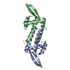
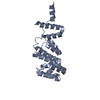
- Assembly
| Deposited unit | 
| ||||||||
|---|---|---|---|---|---|---|---|---|---|
| 1 |
| ||||||||
| 2 | 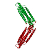
| ||||||||
| Unit cell |
|
-Components
| #1: Protein | CTNNA2 / Alpha N-catenin Mass: 29102.627 Da / Num. of mol.: 1 / Fragment: C-terminal actin-binding domain Source method: isolated from a genetically manipulated source Source: (gene. exp.) Mus musculus (house mouse) / Gene: Catna2, Ctnna2 / Plasmid: pGEX4T1 / Production host:  Escherichia coli (E. coli) / Strain (production host): BL21-CodonPlus / References: UniProt: Q61301 Escherichia coli (E. coli) / Strain (production host): BL21-CodonPlus / References: UniProt: Q61301 | ||||
|---|---|---|---|---|---|
| #2: Chemical | Sulfate  Mass: 96.063 Da / Num. of mol.: 3 / Source method: obtained synthetically / Formula: SO4 Mass: 96.063 Da / Num. of mol.: 3 / Source method: obtained synthetically / Formula: SO4#3: Water | ChemComp-HOH / | Water Mass: 18.015 Da / Num. of mol.: 7 / Source method: isolated from a natural source / Formula: H2O Mass: 18.015 Da / Num. of mol.: 7 / Source method: isolated from a natural source / Formula: H2OSequence details | THE CRYSTALLIZ | |
-Experimental details
-Experiment
| Experiment | Method: X-RAY DIFFRACTION / Number of used crystals: 1 |
|---|
- Sample preparation
| Crystal | Density Matthews: 3.51 Å3/Da / Density % sol: 64.99 % |
|---|---|
| Crystal grow | Temperature: 277 K / Method: vapor diffusion / pH: 7 Details: 0.1M HEPES, pH 7.0, 1.6 M (NH4)2SO4, 0.2 M NaCl,and 0.1M K/Na tartate, vapor diffusion, temperature 277K |
-Data collection
| Diffraction |
| |||||||||||||||||||||||||||||||||||||||||||||||||||||||||||||||||||||||||||||||||||||||||||||||||||||||||||||||||||||||||||||||||||||||||||||||||||
|---|---|---|---|---|---|---|---|---|---|---|---|---|---|---|---|---|---|---|---|---|---|---|---|---|---|---|---|---|---|---|---|---|---|---|---|---|---|---|---|---|---|---|---|---|---|---|---|---|---|---|---|---|---|---|---|---|---|---|---|---|---|---|---|---|---|---|---|---|---|---|---|---|---|---|---|---|---|---|---|---|---|---|---|---|---|---|---|---|---|---|---|---|---|---|---|---|---|---|---|---|---|---|---|---|---|---|---|---|---|---|---|---|---|---|---|---|---|---|---|---|---|---|---|---|---|---|---|---|---|---|---|---|---|---|---|---|---|---|---|---|---|---|---|---|---|---|---|---|
| Diffraction source |
| |||||||||||||||||||||||||||||||||||||||||||||||||||||||||||||||||||||||||||||||||||||||||||||||||||||||||||||||||||||||||||||||||||||||||||||||||||
| Detector |
| |||||||||||||||||||||||||||||||||||||||||||||||||||||||||||||||||||||||||||||||||||||||||||||||||||||||||||||||||||||||||||||||||||||||||||||||||||
| Radiation |
| |||||||||||||||||||||||||||||||||||||||||||||||||||||||||||||||||||||||||||||||||||||||||||||||||||||||||||||||||||||||||||||||||||||||||||||||||||
| Radiation wavelength |
| |||||||||||||||||||||||||||||||||||||||||||||||||||||||||||||||||||||||||||||||||||||||||||||||||||||||||||||||||||||||||||||||||||||||||||||||||||
| Reflection | Resolution: 2.6→50 Å / Num. all: 12400 / Num. obs: 12361 / % possible obs: 99.7 % / Observed criterion σ(F): -3 / Observed criterion σ(I): 0 / Redundancy: 9 % / Biso Wilson estimate: 70.6 Å2 / Rsym value: 0.049 / Χ2: 1.08 / Net I/σ(I): 16.9 | |||||||||||||||||||||||||||||||||||||||||||||||||||||||||||||||||||||||||||||||||||||||||||||||||||||||||||||||||||||||||||||||||||||||||||||||||||
| Reflection shell |
|

- Processing
| Software |
| ||||||||||||||||||||||||||||||||||||||||||||||||||||||||||||||||||||||
|---|---|---|---|---|---|---|---|---|---|---|---|---|---|---|---|---|---|---|---|---|---|---|---|---|---|---|---|---|---|---|---|---|---|---|---|---|---|---|---|---|---|---|---|---|---|---|---|---|---|---|---|---|---|---|---|---|---|---|---|---|---|---|---|---|---|---|---|---|---|---|---|
| Refinement | Method to determine structure: SAD / Resolution: 2.603→39.736 Å / Occupancy max: 1 / Occupancy min: 0.5 / FOM work R set: 0.7918 / SU ML: 0.26 / σ(F): 0 / Phase error: 27.25 / Stereochemistry target values: Engh & Huber
| ||||||||||||||||||||||||||||||||||||||||||||||||||||||||||||||||||||||
| Solvent computation | Shrinkage radii: 0.9 Å / VDW probe radii: 1.2 Å / Solvent model: FLAT BULK SOLVENT MODEL | ||||||||||||||||||||||||||||||||||||||||||||||||||||||||||||||||||||||
| Displacement parameters | Biso max: 120.03 Å2 / Biso mean: 70.8448 Å2 / Biso min: 47.06 Å2 | ||||||||||||||||||||||||||||||||||||||||||||||||||||||||||||||||||||||
| Refine analyze | Luzzati coordinate error obs: 0.26 Å | ||||||||||||||||||||||||||||||||||||||||||||||||||||||||||||||||||||||
| Refinement step | Cycle: LAST / Resolution: 2.603→39.736 Å
| ||||||||||||||||||||||||||||||||||||||||||||||||||||||||||||||||||||||
| Refine LS restraints |
| ||||||||||||||||||||||||||||||||||||||||||||||||||||||||||||||||||||||
| LS refinement shell | Refine-ID: X-RAY DIFFRACTION / Total num. of bins used: 9
|
