 Movie
Movie Controller

[English] 日本語
 Yorodumi
- PDB-3nsu: A Systematic Screen for Protein-Lipid Interactions in Saccharomyc... -
+ Open data

- Basic information
| Entry | Database: PDB / ID: 3nsu | ||||||
|---|---|---|---|---|---|---|---|
| Title | A Systematic Screen for Protein-Lipid Interactions in Saccharomyces cerevisiae | ||||||
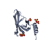
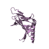
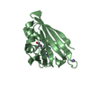
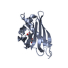
 Components Components | Phosphatidylinositol 4,5-bisphosphate-binding protein SLM1 | ||||||
 Keywords Keywords |  SIGNALING PROTEIN / Pleckstrin homology domain SIGNALING PROTEIN / Pleckstrin homology domain | ||||||
| Function / homology |  Function and homology informationeisosome assembly / sphingolipid binding / establishment or maintenance of actin cytoskeleton polarity / endosomal transport / actin filament bundle assembly / TOR signaling / phosphatidylinositol-4,5-bisphosphate binding / protein localization to plasma membrane / regulation of cell growth / actin cytoskeleton organization ...eisosome assembly / sphingolipid binding / establishment or maintenance of actin cytoskeleton polarity / endosomal transport / actin filament bundle assembly / TOR signaling / phosphatidylinositol-4,5-bisphosphate binding / protein localization to plasma membrane / regulation of cell growth / actin cytoskeleton organization / mitochondrion / identical protein binding / plasma membrane / cytoplasm Function and homology informationeisosome assembly / sphingolipid binding / establishment or maintenance of actin cytoskeleton polarity / endosomal transport / actin filament bundle assembly / TOR signaling / phosphatidylinositol-4,5-bisphosphate binding / protein localization to plasma membrane / regulation of cell growth / actin cytoskeleton organization ...eisosome assembly / sphingolipid binding / establishment or maintenance of actin cytoskeleton polarity / endosomal transport / actin filament bundle assembly / TOR signaling / phosphatidylinositol-4,5-bisphosphate binding / protein localization to plasma membrane / regulation of cell growth / actin cytoskeleton organization / mitochondrion / identical protein binding / plasma membrane / cytoplasmSimilarity search - Function | ||||||
| Biological species |  Saccharomyces cerevisiae (brewer's yeast) Saccharomyces cerevisiae (brewer's yeast) | ||||||
| Method | X-RAY DIFFRACTION / SYNCHROTRON / MOLECULAR REPLACEMENT / Resolution: 2 Å | ||||||
 Authors Authors | Gallego, O. / Fernandez-Tornero, C. / Aguilar-Gurrieri, C. / Muller, C. / Gavin, A.C. | ||||||
 Citation Citation | Journal: Mol. Syst. Biol. / Year: 2010 Title: A systematic screen for protein-lipid interactions in Saccharomyces cerevisiae. Authors: Gallego, O. / Betts, M.J. / Gvozdenovic-Jeremic, J. / Maeda, K. / Matetzki, C. / Aguilar-Gurrieri, C. / Beltran-Alvarez, P. / Bonn, S. / Fernandez-Tornero, C. / Jensen, L.J. / Kuhn, M. / ...Authors: Gallego, O. / Betts, M.J. / Gvozdenovic-Jeremic, J. / Maeda, K. / Matetzki, C. / Aguilar-Gurrieri, C. / Beltran-Alvarez, P. / Bonn, S. / Fernandez-Tornero, C. / Jensen, L.J. / Kuhn, M. / Trott, J. / Rybin, V. / Muller, C.W. / Bork, P. / Kaksonen, M. / Russell, R.B. / Gavin, A.C. | ||||||
| History |
|
- Structure visualization
| Structure viewer | Molecule: MolmilJmol/JSmol |
|---|
- Downloads & links
-Download
| PDBx/mmCIF format | 3nsu.cif.gz | 58.5 KB | Display | PDBx/mmCIF format |
|---|---|---|---|---|
| PDB format | pdb3nsu.ent.gz | 42.1 KB | Display | PDB format |
| PDBx/mmJSON format | 3nsu.json.gz | Tree view | PDBx/mmJSON format | |
| Others |  Other downloads Other downloads |
-Validation report
| Arichive directory | https://data.pdbj.org/pub/pdb/validation_reports/ns/3nsuftp://data.pdbj.org/pub/pdb/validation_reports/ns/3nsu | HTTPS FTP |
|---|
-Related structure data
| Related structure data |  1btkS S: Starting model for refinement |
|---|---|
| Similar structure data |


-Links
 PDBj
PDBj

- Assembly
| Deposited unit | 
| ||||||||
|---|---|---|---|---|---|---|---|---|---|
| 1 | 
| ||||||||
| 2 | 
| ||||||||
| Unit cell |
|
-Components
| #1: Protein | Mass: 13777.617 Da / Num. of mol.: 2 / Fragment: Pleckstrin Homology domain (unp residues 469-583) Source method: isolated from a genetically manipulated source Source: (gene. exp.) Saccharomyces cerevisiae (brewer's yeast)Gene: LIT2, SLM1, YIL105C / Plasmid: pET100-D/Topo / Production host:  Escherichia coli (E. coli) / Strain (production host): BL21 / References: UniProt: P40485 Escherichia coli (E. coli) / Strain (production host): BL21 / References: UniProt: P40485#2: Chemical | ChemComp-SO4 / Sulfate  Mass: 96.063 Da / Num. of mol.: 4 / Source method: obtained synthetically / Formula: SO4 Mass: 96.063 Da / Num. of mol.: 4 / Source method: obtained synthetically / Formula: SO4#3: Water | ChemComp-HOH / | Water Mass: 18.015 Da / Num. of mol.: 107 / Source method: isolated from a natural source / Formula: H2O Mass: 18.015 Da / Num. of mol.: 107 / Source method: isolated from a natural source / Formula: H2O |
|---|
-Experimental details
-Experiment
| Experiment | Method: X-RAY DIFFRACTION / Number of used crystals: 1 |
|---|
- Sample preparation
| Crystal | Density Matthews: 2.05 Å3/Da / Density % sol: 39.95 % |
|---|---|
| Crystal grow | Temperature: 293 K / Method: vapor diffusion, hanging drop / pH: 7.5 Details: 2 M (NH4)2SO4, 2 % PEG 400, 0.1 M Hepes, pH 7.5, VAPOR DIFFUSION, HANGING DROP, temperature 293K |
-Data collection
| Diffraction | Mean temperature: 100 K |
|---|---|
| Diffraction source | Source: SYNCHROTRON / Site: ESRF  / Beamline: ID14-2 / Wavelength: 0.933 Å / Beamline: ID14-2 / Wavelength: 0.933 Å |
| Detector | Type: ADSC QUANTUM 4 / Detector: CCD / Date: Dec 8, 2008 |
| Radiation | Monochromator: Diamond (111), Ge(220) / Protocol: SINGLE WAVELENGTH / Monochromatic (M) / Laue (L): M / Scattering type: x-ray |
| Radiation wavelength | Wavelength: 0.933 Å / Relative weight: 1 |
| Reflection | Resolution: 2→20 Å / Num. obs: 15561 / % possible obs: 97.7 % / Observed criterion σ(F): 0 / Observed criterion σ(I): 0 / Redundancy: 4 % / Biso Wilson estimate: 35.37 Å2 / Rmerge(I) obs: 0.061 / Rsym value: 0.061 / Net I/σ(I): 17.56 |
| Reflection shell | Resolution: 2→2.05 Å / Redundancy: 4 % / Rmerge(I) obs: 0.642 / Mean I/σ(I) obs: 2.19 / Num. unique all: 1136 / Rsym value: 0.642 / % possible all: 99.3 |
- Processing
| Software |
| |||||||||||||||||||||||||||||||||||||||||||||||||
|---|---|---|---|---|---|---|---|---|---|---|---|---|---|---|---|---|---|---|---|---|---|---|---|---|---|---|---|---|---|---|---|---|---|---|---|---|---|---|---|---|---|---|---|---|---|---|---|---|---|---|
| Refinement | Method to determine structure: MOLECULAR REPLACEMENT Starting model: PDB entry 1BTK Resolution: 2→19.824 Å / SU ML: 0.34 / Isotropic thermal model: Isotropic / Cross valid method: THROUGHOUT / σ(F): 2 / Phase error: 27.25 / Stereochemistry target values: ML
| |||||||||||||||||||||||||||||||||||||||||||||||||
| Solvent computation | Shrinkage radii: 0.9 Å / VDW probe radii: 1.11 Å / Solvent model: FLAT BULK SOLVENT MODEL / Bsol: 50.146 Å2 / ksol: 0.359 e/Å3 | |||||||||||||||||||||||||||||||||||||||||||||||||
| Displacement parameters | Biso mean: 30.226 Å2
| |||||||||||||||||||||||||||||||||||||||||||||||||
| Refinement step | Cycle: LAST / Resolution: 2→19.824 Å
| |||||||||||||||||||||||||||||||||||||||||||||||||
| Refine LS restraints |
| |||||||||||||||||||||||||||||||||||||||||||||||||
| LS refinement shell | Refine-ID: X-RAY DIFFRACTION
|
