 Movie
Movie Controller

[English] 日本語
 Yorodumi
- PDB-3zhm: N-terminal domain of the CI repressor from bacteriophage TP901-1 ... -
+ Open data

- Basic information
| Entry | Database: PDB / ID: 3zhm | ||||||
|---|---|---|---|---|---|---|---|
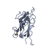
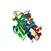
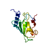
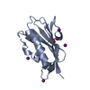
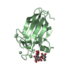
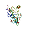
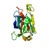
| Title | N-terminal domain of the CI repressor from bacteriophage TP901-1 in complex with the OL2 operator half-site | ||||||
 Components Components |
| ||||||
 Keywords Keywords |  TRANSCRIPTION / TRANSCRIPTION FACTOR / TRANSCRIPTION REGULATION TRANSCRIPTION / TRANSCRIPTION FACTOR / TRANSCRIPTION REGULATION | ||||||
| Function / homology |  Function and homology information Function and homology information | ||||||
| Biological species |  LACTOCOCCUS PHAGE TP901-1 (virus) LACTOCOCCUS PHAGE TP901-1 (virus) | ||||||
| Method | X-RAY DIFFRACTION / SYNCHROTRON / MOLECULAR REPLACEMENT / Resolution: 2.6 Å | ||||||
 Authors Authors | Frandsen, K.H. / Rasmussen, K.K. / Poulsen, J.N. / Lo Leggio, L. | ||||||
 Citation Citation | Journal: Biochemistry / Year: 2013 Title: Binding of the N-Terminal Domain of the Lactococcal Bacteriophage Tp901-1 Ci Repressor to its Target DNA: A Crystallography, Small Angle Scattering, and Nuclear Magnetic Resonance Study. Authors: Frandsen, K.H. / Rasmussen, K.K. / Jensen, M.R. / Hammer, K. / Pedersen, M. / Poulsen, J.N. / Arleth, L. / Lo Leggio, L. | ||||||
| History |
|
- Structure visualization
| Structure viewer | Molecule: MolmilJmol/JSmol |
|---|
- Downloads & links
-Download
| PDBx/mmCIF format | 3zhm.cif.gz | 41.6 KB | Display | PDBx/mmCIF format |
|---|---|---|---|---|
| PDB format | pdb3zhm.ent.gz | 25.5 KB | Display | PDB format |
| PDBx/mmJSON format | 3zhm.json.gz | Tree view | PDBx/mmJSON format | |
| Others |  Other downloads Other downloads |
-Validation report
| Arichive directory | https://data.pdbj.org/pub/pdb/validation_reports/zh/3zhmftp://data.pdbj.org/pub/pdb/validation_reports/zh/3zhm | HTTPS FTP |
|---|
-Related structure data
| Related structure data |  3zhiC  2r1jS C: citing same article ( S: Starting model for refinement |
|---|---|
| Similar structure data |


-Links
 PDBj
PDBj


- Assembly
| Deposited unit | 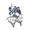
| ||||||||
|---|---|---|---|---|---|---|---|---|---|
| 1 |
| ||||||||
| Unit cell |
|
-Components
| #1: Protein | Mass: 9105.513 Da / Num. of mol.: 1 / Fragment: RESIDUES 2-74 Source method: isolated from a genetically manipulated source Source: (gene. exp.) LACTOCOCCUS PHAGE TP901-1 (virus) / Plasmid: PQE-70 / Production host:  ESCHERICHIA COLI (E. coli) / Strain (production host): M15 / References: UniProt: O48503 ESCHERICHIA COLI (E. coli) / Strain (production host): M15 / References: UniProt: O48503 |
|---|---|
| #2: DNA chain | Mass: 5515.591 Da / Num. of mol.: 1 / Source method: obtained synthetically / Source: (synth.) LACTOCOCCUS PHAGE TP901-1 (virus) |
| #3: DNA chain | Mass: 5515.591 Da / Num. of mol.: 1 / Source method: obtained synthetically / Source: (synth.) LACTOCOCCUS PHAGE TP901-1 (virus) |
| #4: Water | ChemComp-HOH / Water Mass: 18.015 Da / Num. of mol.: 56 / Source method: isolated from a natural source / Formula: H2O Mass: 18.015 Da / Num. of mol.: 56 / Source method: isolated from a natural source / Formula: H2O |
| Sequence details | OLIGONUCLE |
-Experimental details
-Experiment
| Experiment | Method: X-RAY DIFFRACTION |
|---|
- Sample preparation
| Crystal | Density Matthews: 2.19 Å3/Da / Density % sol: 43.45 % / Description: NONE |
|---|---|
| Crystal grow | pH: 7 / Details: pH 7 |
-Data collection
| Diffraction | Mean temperature: 100 K |
|---|---|
| Diffraction source | Source: SYNCHROTRON / Site: ESRF  / Beamline: ID23-2 / Wavelength: 0.87 / Beamline: ID23-2 / Wavelength: 0.87 |
| Detector | Type: ADSC CCD / Detector: CCD |
| Radiation | Protocol: SINGLE WAVELENGTH / Monochromatic (M) / Laue (L): M / Scattering type: x-ray |
| Radiation wavelength | Wavelength: 0.87 Å / Relative weight: 1 |
| Reflection | Resolution: 2.6→33.91 Å / Num. obs: 4365 / % possible obs: 100 % / Observed criterion σ(I): 1.36 / Redundancy: 4.4 % / Biso Wilson estimate: 22.41 Å2 / Rmerge(I) obs: 0.16 / Net I/σ(I): 9 |
| Reflection shell | Resolution: 2.6→2.74 Å / Redundancy: 4.6 % / Rmerge(I) obs: 0.49 / Mean I/σ(I) obs: 4.7 / % possible all: 100 |
- Processing
| Software |
| ||||||||||||||||||||||||||||
|---|---|---|---|---|---|---|---|---|---|---|---|---|---|---|---|---|---|---|---|---|---|---|---|---|---|---|---|---|---|
| Refinement | Method to determine structure: MOLECULAR REPLACEMENT Starting model: PDB ENTRY 2R1J Resolution: 2.6→33.91 Å / SU ML: 0.17 / σ(F): 1.36 / Phase error: 17.9 / Stereochemistry target values: ML
| ||||||||||||||||||||||||||||
| Solvent computation | Shrinkage radii: 0.9 Å / VDW probe radii: 1.11 Å / Solvent model: FLAT BULK SOLVENT MODEL | ||||||||||||||||||||||||||||
| Refinement step | Cycle: LAST / Resolution: 2.6→33.91 Å
| ||||||||||||||||||||||||||||
| Refine LS restraints |
| ||||||||||||||||||||||||||||
| LS refinement shell |
|
