 Movie
Movie Controller

[English] 日本語
 Yorodumi
- PDB-5dil: Crystal structure of the effector domain of the NS1 protein from ... -
+ Open data

- Basic information
| Entry | Database: PDB / ID: 5dil | ||||||
|---|---|---|---|---|---|---|---|
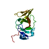
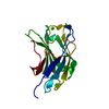
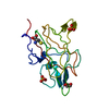
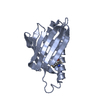
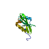
| Title | Crystal structure of the effector domain of the NS1 protein from influenza virus B | ||||||
 Components Components | Non-structural protein 1 | ||||||
 Keywords Keywords |  RNA-binding Protein / Viral Protein / effector domain / RNA binding RNA-binding Protein / Viral Protein / effector domain / RNA binding | ||||||
| Function / homology |  Function and homology information Function and homology informationsymbiont-mediated suppression of host PKR/eIFalpha signaling / symbiont-mediated suppression of host ISG15-protein conjugation / host cell cytoplasm / symbiont-mediated suppression of host type I interferon-mediated signaling pathway / host cell nucleus / RNA bindingSimilarity search - Function | ||||||
| Biological species |  Influenza B virus Influenza B virus | ||||||
| Method | X-RAY DIFFRACTION / SAD / Resolution: 2.01 Å | ||||||
 Authors Authors | Guan, R. / Hamilton, K. / Ma, L. / Montelione, G.T. | ||||||
| Funding support |  United States, 1items United States, 1items
| ||||||
 Citation Citation | Journal: Structure / Year: 2016 Title: A Second RNA-Binding Site in the NS1 Protein of Influenza B Virus. Authors: Ma, L.C. / Guan, R. / Hamilton, K. / Aramini, J.M. / Mao, L. / Wang, S. / Krug, R.M. / Montelione, G.T. | ||||||
| History |
|
- Structure visualization
| Structure viewer | Molecule: MolmilJmol/JSmol |
|---|
- Downloads & links
-Download
| PDBx/mmCIF format | 5dil.cif.gz | 68.4 KB | Display | PDBx/mmCIF format |
|---|---|---|---|---|
| PDB format | pdb5dil.ent.gz | 53.7 KB | Display | PDB format |
| PDBx/mmJSON format | 5dil.json.gz | Tree view | PDBx/mmJSON format | |
| Others |  Other downloads Other downloads |
-Validation report
| Arichive directory | https://data.pdbj.org/pub/pdb/validation_reports/di/5dilftp://data.pdbj.org/pub/pdb/validation_reports/di/5dil | HTTPS FTP |
|---|
-Related structure data
| Similar structure data |
|---|


-Links
 PDBj
PDBj- Assembly
| Deposited unit | 
| ||||||||
|---|---|---|---|---|---|---|---|---|---|
| 1 | 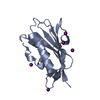
| ||||||||
| 2 | 
| ||||||||
| Unit cell |
|
-Components
| #1: Protein | Mass: 16010.823 Da / Num. of mol.: 2 / Fragment: UNP residues 141-281 Source method: isolated from a genetically manipulated source Source: (gene. exp.) Influenza B virus (B/Singapore/DSO_090134/2004)Strain: B/Singapore/DSO_090134/2004 / Gene: NS1 / Production host:  Escherichia coli (E. coli) / References: UniProt: X2C382 Escherichia coli (E. coli) / References: UniProt: X2C382#2: Chemical | ChemComp-IOD / Iodide  Mass: 126.904 Da / Num. of mol.: 13 / Source method: obtained synthetically / Formula: I Mass: 126.904 Da / Num. of mol.: 13 / Source method: obtained synthetically / Formula: I#3: Water | ChemComp-HOH / | Water Mass: 18.015 Da / Num. of mol.: 200 / Source method: isolated from a natural source / Formula: H2O Mass: 18.015 Da / Num. of mol.: 200 / Source method: isolated from a natural source / Formula: H2O |
|---|
-Experimental details
-Experiment
| Experiment | Method: X-RAY DIFFRACTION |
|---|
- Sample preparation
| Crystal | Density Matthews: 2.05 Å3/Da / Density % sol: 39.93 % |
|---|---|
| Crystal grow | Temperature: 277 K / Method: vapor diffusion, hanging drop / pH: 5 / Details: 0.2M KI 20% PEG3350 |
-Data collection
| Diffraction | Mean temperature: 100 K |
|---|---|
| Diffraction source | Source: ROTATING ANODE / Type: RIGAKU / Wavelength: 1.5418 Å |
| Detector | Type: RIGAKU RAXIS IV++ / Detector: IMAGE PLATE / Date: Apr 30, 2013 |
| Radiation | Protocol: SINGLE WAVELENGTH / Monochromatic (M) / Laue (L): M / Scattering type: x-ray |
| Radiation wavelength | Wavelength: 1.5418 Å / Relative weight: 1 |
| Reflection | Resolution: 2.01→46.22 Å / Num. obs: 16041 / % possible obs: 95 % / Redundancy: 3.9 % / Rmerge(I) obs: 0.084 / Net I/σ(I): 12.22 |
- Processing
| Software |
| ||||||||||||||||||||||||||||||||||||||||||||||||||||||||||||||||||||||||||||||||||||||||||||||||||||||||||||||||||||||||||||||||||||||||||||||||||||||||||||||||||||||||
|---|---|---|---|---|---|---|---|---|---|---|---|---|---|---|---|---|---|---|---|---|---|---|---|---|---|---|---|---|---|---|---|---|---|---|---|---|---|---|---|---|---|---|---|---|---|---|---|---|---|---|---|---|---|---|---|---|---|---|---|---|---|---|---|---|---|---|---|---|---|---|---|---|---|---|---|---|---|---|---|---|---|---|---|---|---|---|---|---|---|---|---|---|---|---|---|---|---|---|---|---|---|---|---|---|---|---|---|---|---|---|---|---|---|---|---|---|---|---|---|---|---|---|---|---|---|---|---|---|---|---|---|---|---|---|---|---|---|---|---|---|---|---|---|---|---|---|---|---|---|---|---|---|---|---|---|---|---|---|---|---|---|---|---|---|---|---|---|---|---|
| Refinement | Method to determine structure: SAD / Resolution: 2.01→28.079 Å / SU ML: 0.25 / Cross valid method: FREE R-VALUE / σ(F): 1.98 / Phase error: 26.3 / Stereochemistry target values: ML
| ||||||||||||||||||||||||||||||||||||||||||||||||||||||||||||||||||||||||||||||||||||||||||||||||||||||||||||||||||||||||||||||||||||||||||||||||||||||||||||||||||||||||
| Solvent computation | Shrinkage radii: 0.9 Å / VDW probe radii: 1.11 Å / Solvent model: FLAT BULK SOLVENT MODEL | ||||||||||||||||||||||||||||||||||||||||||||||||||||||||||||||||||||||||||||||||||||||||||||||||||||||||||||||||||||||||||||||||||||||||||||||||||||||||||||||||||||||||
| Refinement step | Cycle: LAST / Resolution: 2.01→28.079 Å
| ||||||||||||||||||||||||||||||||||||||||||||||||||||||||||||||||||||||||||||||||||||||||||||||||||||||||||||||||||||||||||||||||||||||||||||||||||||||||||||||||||||||||
| Refine LS restraints |
| ||||||||||||||||||||||||||||||||||||||||||||||||||||||||||||||||||||||||||||||||||||||||||||||||||||||||||||||||||||||||||||||||||||||||||||||||||||||||||||||||||||||||
| LS refinement shell |
|
